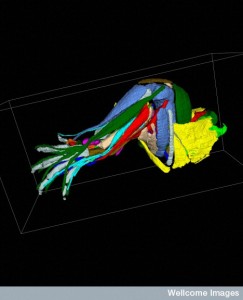
B0007551 Muscles and tendons of a mouse hindlimb
Credit: NIMR, MRC. Wellcome Images
images@wellcome.ac.uk
http://wellcomeimages.org
This image shows the structue and positioning of the muscles and tendons present in the hindlimb of a 14.5 days post coitum (dpc) mouse embryo. The different muscles are colour coded for definition. This image is part of a wider limb atlas project created to identify the structure of bones, muscles and tendons in the developing limb. The limb atlas has been developed by scientists at the NIMR, MRC London.
Optical Projection Tomography
Published: –
Copyrighted work available under Creative Commons by-nc-nd 4.0, see http://wellcomeimages.org/indexplus/page/Prices.html
