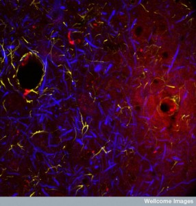
B0002694 Haemorrhaging blood vessel in the brain
Credit: MRC Toxicology Unit. Wellcome Images
images@wellcome.ac.uk
http://wellcomeimages.org
Confocal image showing a haemorrhaging blood
vessel in the brain. In normal circumstances
only selective small molecules are able to cross
into the surrounding tissue but in many cases of
injury and disease the barriers break down and
blood can be released causing tissue damage. In
this image blood plasma (red) is leaking into the
surrounding tissue. Neurones (blue) and glial
cells (yellow/green) can be seen in the adjacent
tissue but are damaged and therefore less
prevalent in the area of the leak.
Confocal micrograph
Published: –
Copyrighted work available under Creative Commons by-nc-nd 4.0, see http://wellcomeimages.org/indexplus/page/Prices.html
