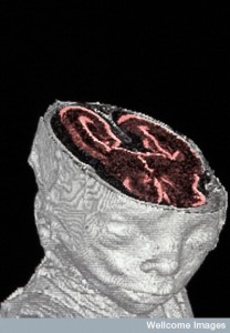
B0001014 Foetal MRI scan
Credit: Nathan Jeffery. Wellcome Images
images@wellcome.ac.uk
http://wellcomeimages.org
A high resolution magnetic resonance imaging
(HRMRI) scan of a miscarried 20-week old human
foetus, showing the head and a cross-section of
the brain. This technique uses a magnetic field to
induce the hydrogen nuclei in tissues to absorb
and emit radio waves, producing an image which
allows internal structures to be seen without
disturbing the actual specimen.
Published: –
Copyrighted work available under Creative Commons by-nc-nd 4.0, see http://wellcomeimages.org/indexplus/page/Prices.html
