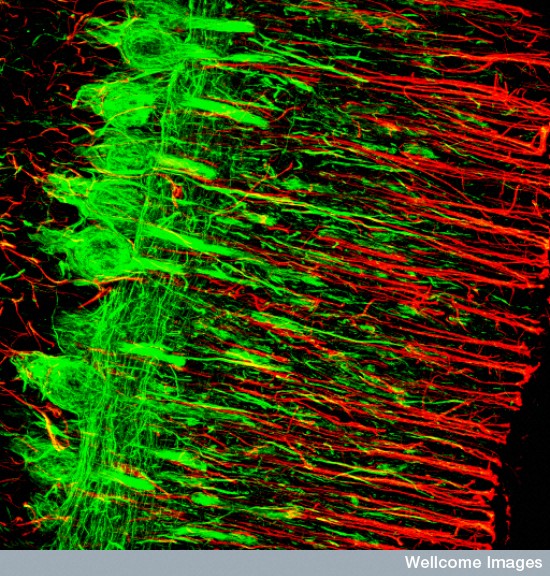
B0001869 Purkinje cells and radial glia
Credit: MRC Toxicology Unit. Wellcome Images
images@wellcome.ac.uk
http://wellcomeimages.org
Confocal microscopic image of Purkinje cells and
radial glia in the cerebellum. The Purkinje cells
stain green with an antibody to neurofilament and
the glial cells appear red, detecting an antibody
to Glila fibrillary Acidic Protein (GFAP). The
cell bodies of the Purkinje cells show dense
neurofilaments in a row to the left of the
picture. The radial Glia of Bergmann intercalate
with the Purkinje cell processes.
Confocal micrograph
Published: –
Copyrighted work available under Creative Commons by-nc-nd 4.0, see http://wellcomeimages.org/indexplus/page/Prices.html