B0008436 Cerebellar circuitry
Credit: Praneeth Namburi. Wellcome Images
images@wellcome.ac.uk
http://wellcomeimages.org
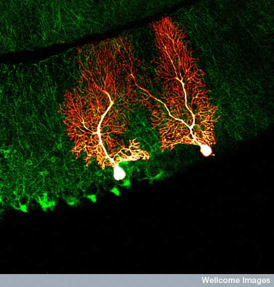
This image was produced using two photon microscopy, which allows fluorescent imaging of living tissue. The image shows the structure of cerebellar circuitry in a transgenic mouse. Interneurons in the cerebellum that express neuronal NOS (Nitiric Oxide Synthetase) enzyme are shown in green and two of the Purkinje cells in the Purkinje cell layer are shown in red.
Fluorescence microscopy
2011 Published: –
Copyrighted work available under Creative Commons by-nc-nd 4.0, see http://wellcomeimages.org/indexplus/page/Prices.html