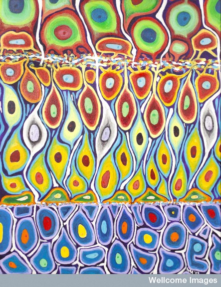
B0009505 Cell fates in zebrafish retina, acrylic painting
Credit: Prof. Bill Harris. Wellcome Images
images@wellcome.ac.uk
http://wellcomeimages.org
Artist’s impression of the cellular structure of part of a developing zebrafish retina. The retina is almost fully developed so all the cells have been born and have almost finished moving into one of three layers depicted here. The top layer is the ganglion cell layer which contains retinal ganglion cells. The middle layer (inner nuclear layer) contains horizontal cells (green), bipolar cells (yellow), Mueller cells (white) and amacrine cells (red). The cells in the bottom layer are cells of the outer nuclear layer (rods and cones). This acrylic painting was inspired by multi-coloured fluorescent proteins which are used to label different types of cells and different compartments inside a cell (e.g. nucleus, membrane, cytoplasm). This gives an instantaneous view of the spectrum of cell fates within a neural tissue.
Artwork
2013 Published: –
Copyrighted work available under Creative Commons by-nc-nd 4.0, see http://wellcomeimages.org/indexplus/page/Prices.html