B0002158 Alzheimer’s brain showing plaque & microglia
Credit: Philippe Gasque. Wellcome Images
images@wellcome.ac.uk
http://wellcomeimages.org
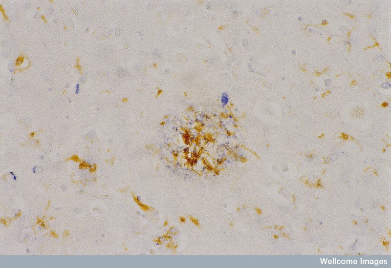
A section through the temporal cortex of a
brain with Alzheimer’s disease. The nuclei are
weakly stained blue with haemotoxylin and the
amyloid proteins of the neuritic plaque are
stained in dark blue. The brown cells are
microglia trying to clear the amyloid fibrils by
phagocytosis. Microglia are the macrophages of
the central nervous system and although they are
trying to clear the neuritic plaques they can also
produce molecules toxic to the surrounding neural
cells.
Published: –
Copyrighted work available under Creative Commons by-nc-nd 4.0, see http://wellcomeimages.org/indexplus/page/Prices.html