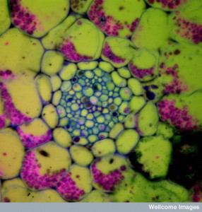
B0005175 Section of Neottia root, showing mycorrhiza, fluorescent
Credit: Jim Haseloff. Wellcome Images
images@wellcome.ac.uk
http://wellcomeimages.org
A transverse section through a root of bird’s nest orchid (Neottia sp), viewed using a confocal microscope. Cells are labelled as a result of staining with a coloured fluorescent probe and autofluorescence. The image shows the presence of fungal threads (brown/blue) in the cortex. These constitute an endomycorrhiza, a symbiotic relationship between plant and fungus in which the fungus penetrates the root tissues and contributes to its nutrition. Many of the cortex cells contain starch grains (pink) for food storage.
Confocal micrograph
Published: –
Copyrighted work available under Creative Commons by-nc-nd 4.0, see http://wellcomeimages.org/indexplus/page/Prices.html
