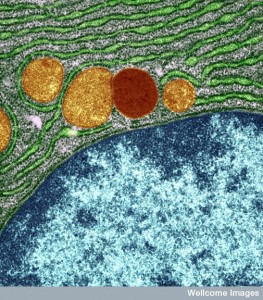
B0004343 Organelles in a pancreas cell
Credit: University of Edinburgh. Wellcome Images
images@wellcome.ac.uk
http://wellcomeimages.org
Colour-enhanced electron micrograph of part of a pancreas cell showing the nucleus in blue, mitochondria in orange, a lysosome in red and rough endoplasmic reticulum in green. A nuclear pore is also visible in the nuclear membrane towards the right-hand end.
The horizontal field width of the sample is 2.9 micrometres.
Transmission electron micrograph
1980-2000 Published: –
Copyrighted work available under Creative Commons by-nc-nd 4.0, see http://wellcomeimages.org/indexplus/page/Prices.html
