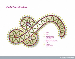
B0009905 Ebola virus structure, illustration
Credit: Pete Jeffs. Wellcome Images
images@wellcome.ac.uk
http://wellcomeimages.org
Illustration of the structure of a single ebola virus particle. The ebola virus belongs to the Filoviridae family of viruses and causes ebola virus disease (EVD) or ebola haemorrhagic fever in humans. Symptoms of this often fatal illness include sudden onset of fever, headache, joint and muscle pain, sore throat and intense muscle weakness followed by diarrhoea, vomiting, stomach pain, impaired kidney and liver function, and in some cases internal and external bleeding. Ebola virus disease first appeared during outbreaks in Africa in the mid-1970s. The virus spreads between humans through direct contact with infected blood, secretions and organs, or with surfaces or bedding already contaminated with these fluids.
Ebola virus particles (virions) are cylindrical or tubular in shape and are made up of three main components: viral envelope, matrix, and nucleocapsid. Ebola virus particles can be up to 1000 nm long and 80 nm in diameter. They have glycoproteins projecting from the surface in 7 – 10 nm spikes (yellow). Viral proteins VP40 (red) and VP24 (orange) are found between the envelope and the nucleocapsid. The virus carries a negative sense RNA genome which is about 18 – 19 Kb long, and is helically wound (green).
Illustration
2014 Published: –
Copyrighted work available under Creative Commons by-nc-nd 4.0, see http://wellcomeimages.org/indexplus/page/Prices.html
