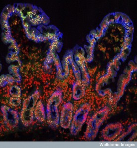
B0006676 Villi from the small intestine
Credit: S. Schuller. Wellcome Images
images@wellcome.ac.uk
http://wellcomeimages.org
Villi from the human small intestine. The finger-like shape increases the surface area of the intestine, improving the efficiency of food absorption. The intestinal glands or crypts can be seen below the villi. The green stain highlights the nucleoli of the rapidly growing epithelial layer of both the villi and the crypts. All the nuclei are stained red and the epithelial cell membranes, blue.
Confocal micrograph
2007 Published: –
Copyrighted work available under Creative Commons by-nc-nd 4.0, see http://wellcomeimages.org/indexplus/page/Prices.html
