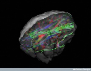
B0008398 White matter fibres of the uncinate fasciculus
Credit: Christopher Whelan, Royal College of Surgeons in Ireland. Wellcome Images
images@wellcome.ac.uk
http://wellcomeimages.org
This image shows an inferior (bottom) view of the white matter fibers of the uncinate fasciculus (connector of the temporal and frontal brain regions) in a neurologically healthy individual, produced using Diffusion Tensor Imaging (DTI). This image was constructed in order to advance the understanding of the neural connections involved in the secondarily generalisation of temporal lobe seizures.
Secondarily-generalized tonic-clonic seizures (SGTCS) frequently referred to as grand mal attacks are the most dangerous types of epileptic seizure. They involve a bilateral, symmetrical surge of neuronal activity which leads to both a tonic seizure phase (principally involving rigidity, crying out, tongue biting, apnea, cyanosis and an increase in blood rate) and a clonic phase in which the patient falls and develops intermittent clonic movements typically involving all four limbs featuring brief interludes of muscle relaxation. The precise mechanism by which these seizures spread from the site of onset to the rest of the brain is still unclear.
In order to understand a potential mechanism of this seizure spread in temporal lobe epilepsy (TLE), diffusion sensor imaging was used to compare the white matter tracts in the uncinate fasciculus in healthy individuals, to those suffering from SGTCS and to patients who remained seizure-free for at least two years.
Diffusion tensor imaging (DTI)
21/12/2011 Published: –
Copyrighted work available under Creative Commons by-nc-nd 4.0, see http://wellcomeimages.org/indexplus/page/Prices.html
