B0000104 3T3 fibroblast cell
Credit: Dr David Becker. Wellcome Images
images@wellcome.ac.uk
http://wellcomeimages.org
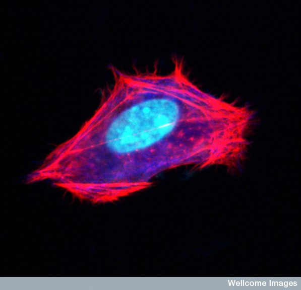
Confocal microscope image of a 3T3 fibroblast cell
before it divides in culture. The nucleus has been
stained blue, whilst the actin microfilaments
which form part of the cytoskeleton are stained
red. The cytoskeleton forms the internal framework
of the cell, giving it shape.
Credit: D. Becker & K. Whitley
Confocal micrograph
Published: –
Copyrighted work available under Creative Commons by-nc-nd 4.0, see http://wellcomeimages.org/indexplus/page/Prices.html