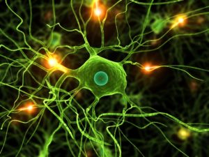
 As both unique immune cells and unique brain cells that constantly change shape and have numerous different functions, are microglia the most intelligent brain cells? Microglia travel independently, not attached to any structure, constantly circling a territory with extended arms repeatedly tapping all axons, dendrites and synapses looking to detect any suboptimal functioning. Their constant surveillance of the brain protects against any microbe invaders, demyelination, trauma and cancerous or defective cells.
But, this is just the beginning of their amazing functions. They communicate with complex wireless signaling to neurons and astrocytes, determining how many brains cells are needed and when to eliminate a synapse—eating the defective or unused synapses. They determine 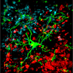 the rate of production of new cells from stem cells and the rate of total brain growth in the fetus—eating excessive stem cells. They morph into many different shapes and types of immune cells to fight a multitude of different invaders. How can we not say that these are intelligent cells?
Microglia have as many functions, if not more, than T cells. A previous post showed the great intelligence of T cells—the masters of immune cells. The independent intelligent T cells travel through the entire body, finding invaders, trauma and cancer. They are critical for cognitive brain function sending signals to brain cells. When travelling in the CSF, T cells are able to send signals to brain cells to increase cognition and suppress other immune cells to avoid unnecessary inflammation in the brain. Then, when they detect a microbe antigen, they switch gears, and unleash an inflammation response while sending signals to the brain that cause a decrease in cognition–stimulating the “sick feeling” which increases rest during illness. 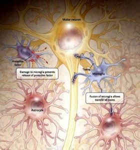
Microglia are the resident immune cells in the brain, but do so much more than just immune surveillance. They help establish neuronal networks in the fetus. In the adult, microglia are actively involved in pruning neurons that are established and nourished by astrocytes. They produce signals that nourish and stimulate neuronal growth and axon migration. The neuron is the master of information processing and transfer. The astrocyte provides nourishment and blood flow to the neurons and participates in the information transfer with its own system; astrocytes, also, establish a protective structure for the neuron and control blood flow to the region. But, it is the microglia who travel throughout the brain participating in surveillance, stimulation, cleanup, and maintenance tasks while communicating with all other cells.
Where Do Microglia Come From?
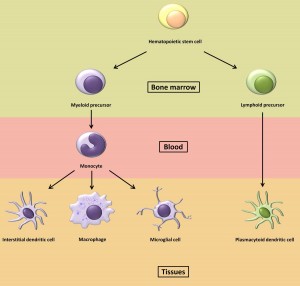 Because they are so unique even among immune cells, there has been a question about their lineage. Recently, it was discovered that they come from a different source in the fetus than the neuron, astrocyte, and oligodendrocyte. They are, in fact, a close cousin of the macrophage, a white blood cell whose major function is gobbling up invaders.
Microglia arise from mesodermal tissue in the yolk sac on day 9 in humans and then travel through the blood to the brain where they stay. In the brain, microglia replenish their own stock—making fewer new cells when they are quietly circling and tapping synapses, and more when they are fighting invaders or debris. They are a stable independent community of cells patrolling their individual territory. 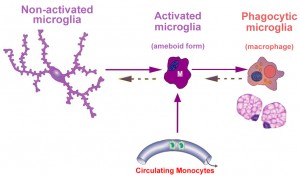
Ordinary macrophages originate from the bone marrow and travel through the blood to different regions of the body as needed. Strangely, there exist special subpopulations of microglia that are, also, from bone marrow. These macrophage cells live in the perivascular region. If a large number of microglia are eliminated in a battle with microbes, then macrophages from outside the brain take their place. These substitute cells gradually, over a period of 8 months, accommodate to the brain, but never learn all of the functions of microglia.
Microglia are much more highly regulated than ordinary macrophages, with specific territories and very exact immune responses like a T cell.
Microglia in Many Shapes and Roles
Microglia are as common as neurons in the brain. It took many years to figure out that a wide variety of different shaped cells in the brain were actually all the same. Microglia 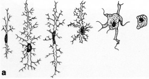 have many different shapes for different functions and therefore have been difficult to study. No one realized that the large blob-like macrophages were the same as star-like cells tapping synapses. Microglia are found near any inflammation, trauma, autoimmune disease, and cancer. Under normal circumstances they are present evenly throughout the brain.
From a spider like resting state, microglia become a big round blob during attacks on microbes as a macrophage. Microglia are very sensitive, part of the immune system and  the brain. With the slightest injury to a nerve or a microbe, microglia suddenly become active and often change shape.
Among the many different shapes are the amoeboid cell crawling to find debris; the quiet “ramified” cell where the arms are moving; the activated cell; a blob fighting and eating microbes; a long rod fighting syphilis; a large granular corpuscle filled with debris from the fight; a perivascular cell repairing vessel walls; and a cell in the juxta-vascular basal lamina. 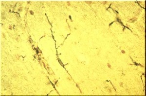
Before modern cellular imaging, it appeared that microglia were very quiet with many long arms. Recently, they have been observed moving steadily like amoeba in their particular territory with many very long arms sticking through the astrocyte and neuronal matrix, wrapping around synapses, axons and dendrites and constantly while moving studying the terrain.
The arms appear to grow, shrink and then regrow. Microglia are, by far, the most active cells in the brain.
Microglia are the most individualistic cell in the brain and function alone, although in constant wireless communication with both immune and brain cells, like the T cell. They have specific territory and don’t tread on each other’s region of surveillance unless there is danger when they swing into action and can go anywhere.
Microglia Respond to Many Different Threats
 Microglia respond to many different stimuli. Surprisingly, they respond to distant threats in the nervous system as well as local. The response can occur within minutes and last for days or longer and can occur in many different ways. They are the only cells that are not connected in the brain and respond to a wide variety of wireless cytokine signals.
When damage occurs in the brain from trauma or infection, microglia are the first responders starting to clean up the debris. They were first discovered near injury, looking like amoebae crawling to the injury site and eating dead microbes and neurons along the way—making room for healing. 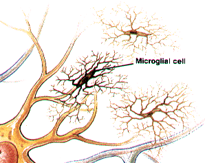
Microglia respond to internal and external threats to the entire body. These responses are sometimes related to specific brain regions, but sometimes not. Stress increases microglia in the brain’s stress regions (see post: social perception affects immune response). They are general protectors of the spinal cord and brain. Those that respond to pain are not just inflammatory, but, appear to be involved in higher level coordination of defense with many other types of cells.
Microglia don’t usually use the antigen process. But, when they respond as an immune cell they do use MHC antigens as do lymphocytes. Human microglia has one very specific gene that sends a signal, which suppresses inflammatory reactions in the brain.
Constant Wireless Communication – Signaling
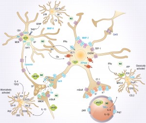 Like T cells, microglia grow a wide range of receptors that can pick up a large number of cytokine signals from other immune and brain cells that are circulating in blood and tissue. Humans, likewise, build their own types of receptors—radios, TV, cellular phones, and laptops—that receive the many wireless signals floating in the air. Both microglia and humans also return signals—microglia use cytokines and neurotransmitters, while humans send radio waves, text and voice messages and emails.
Microglia, using receptors and signals, are in constant communication with neurons, astrocytes and immune cells. Microglia receive neuronal signals of neurotransmitters and send signals mediating neuronal activity. Neurons send signals that stimulate microglia inflammatory behavior and synapse and apoptosis pruning 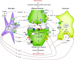 behavior. These microglia behaviors are gradually trained in response to their relationship with neurons.
Signals can promote or fight inflammation and can use MHC receptors when necessary. Microglia are the only cell in the brain that has a complement receptor.
Many very specific signals are secreted in specific disease states such as pain, autoimmune disease, trauma, prion disease, Alzheimer’s and other degenerative brain disease. Signals include cytokines, neurotransmitters, chemokines (specific cytokines that tell cells where to travel) and proteases that alter the extracellular matrix (see post).
Microglia Care for Neurons
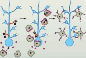 With special dyes, microglia have been observed constantly moving in a specific circular region of about 80 micrometers every several hours with their long arms tapping and examining every part of the region.
One of their roles is to find synapses marked by complement that need to be eliminated. But, their role in eating defective or poorly used synapses is, also, regulated by a back and forth signaling communication with neurons and astrocytes. Healthy neurons secrete signals to tell the microglia not to attack the synapse, while complement can be attracted by the signals from the neurons themselves. Astrocytes send signals to draw microglia to the pre and post synaptic cells of specific synapses. Somehow, microglia know how many new neurons are needed and stop stem cells from making too many. This week research found that astrocytes are active, along with microglia, in pruning some of the unused synapses. 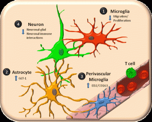
During normal function, the microglia are active in stimulating new neurons and new neuronal connections and providing guidance for long travelling cells and axons. When neurons are overgrown from lack of use and would create problems if not pruned, the microglia are the cells that eat back the unnecessary growth. When defective, neurons can trigger apoptosis and kill themselves; it is the microglia that clean up the debris.
Microglia are involved in memory production. They produce nutrients for neurons that stimulate new dendrite and axon budding. They are actively involved in signals and nutrients for the developing synapse along with astrocytes. One of the most active areas for microglia is the hippocampus the center of memory.
Microglia Necessary to Build the Fetal Brain
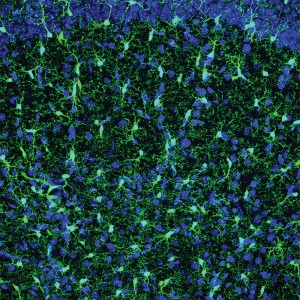 When the fetal brain rapidly grows at the fantastic rate of 250,000 new neurons each second during the last month of pregnancy, the connections that are not used by experience are eliminated by microglia. During childhood and adolescence many connections are made and, also, pruned by experience. It is microglia that mold the adult brain through this process.
Microglia control the number of stem cells that determine how large the cortex grows in the fetal brain. They hover near the neural stem cells in the developing brain and when activated there is less stem cell activity. They are the inhibitory cells of the growing brain. There are specific regions where neurons grow in the fetus as the brain is built. These proliferative zones are filled with microglia. As the brain is built, the number of microglia changes and they evenly space themselves in their individual territories throughout the entire brain.
Microglia Critical for Neuroplasticity
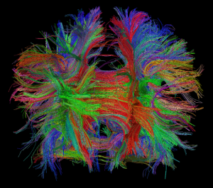 Microglia are critical for neuroplasticity and for the development of synapses. Many experiments show the dramatic rewiring of axon/dendrite connections during any changing circumstances. One experiment with mice in light and dark conditions showed that during several days of dark dendritic spines connecting to axons shrivel and then grow again when there is stimulation with light.
When the dendrites are shrinking, microglia are present to eliminate the unnecessary ones. When the dendrites are growing, the microglia move away and continue their surveillance. The microglia are removing the unused, weak and useless connections to allow the new connections to be vigorous.
Microglia, also, secrete many special nerve factors that stimulate more neurons, survival of neurons, and more connections. They also stimulate myelination as well as blood growth. Eating of synapses can actually stimulate regeneration of neurons.
Microglia and Disease
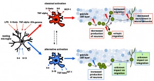 Microglia are more active in many diseases such as Alzheimer’s, Autism, multiple sclerosis, syphilis, HIV, HSV and depression. But, it is not certain if they are helping or hurting.
They may be involved in the shrunken brain regions of schizophrenia, possibly from fetal infections. While fighting syphilis and some other infections, microglia can take the form of “rod cells.” When innate immunity is triggered, microglia change and become part of the response, presenting antigens to lymphocytes and T cells. They function as a dendritic cell, a macrophage and other immune cell types. 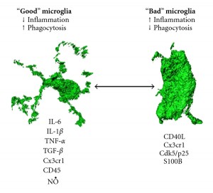
Microglia can become cytotoxic and aggressive. In Rett syndrome they damage dendrites and synapses releasing glutamate. HIV virus infects microglia as their main target in the brain. Microglia infected with HIV release signals that are toxic to neurons helping to cause HIV dementia. This also appears to occur in HSV encephalitis, where microglia can be infected for a year after antiviral treatment.
Microglia in Alzheimer’s
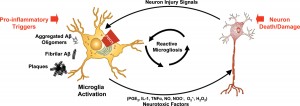 In Alzheimer’s microglia can be both helpful and harmful. Microglia synthesize amyloid precursor protein (APP) in response to injury from too much neuronal excitation. When APP in the neuronal membrane is abnormally cut it produces amyloid plaques, which stimulate many cytokines, toxins, and nitric oxide that helps to kill neurons. Microglia, also, accumulate with tau pathology and precede degeneration. More often microglia are helpful, not harming other neurons by limiting amyloid beta.
Microglia Increase Pain
Microglia are critical to many types of pain.  In neuropathic pain, neurons in the dorsal horn become hyper excitable. In these painful states microglia are activated through a wide variety of signals from damaged neurons that are picked up by receptors on microglia. Microglia are activated by these signals and they secrete special signals causing neuroplastic increase in excitability and inflammation in pain fibers. Signals from microglia, interleukin-1beta and tumour necrosis factor- alpha, activate signals that are toxic to nerve cells, even causing cell death.
Dental inflammation stimulates microglia in the trigeminal subnucleus caudalis. After heart attack microglia are in the hypothalamic parventricular nucleus causing pain. Microglia signals are also important in morphine tolerance.
Microglia and Prions
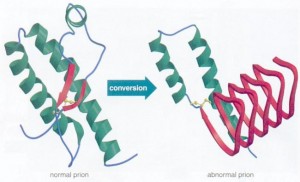 Microglia try to eat the prions (see post), but instead often transport them into the brain. They can either help the situation or hurt. Brains with prion disease have many microglia present. For microglia to help against prions they must have effective signaling with astrocytes.
In the battle with prions microglia alter their cell type from M1, which fights inflammation to M2, which causes inflammation. M2 spreads the prion further. As prion accumulates in the brain, many cytokines are stimulated, which increase inflammation—IL-1, TNF and IL-6. But, at the same times anti-inflammatory cytokines are also increased—IL-4 and IL-10 in CSF. Also, anti nuclear factor, NF-kappa-beta is stimulated, which causes neuronal apoptosis (cell death).
Microglia and Abnormal Human Behavior
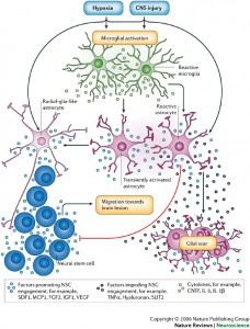 An alteration in the human hoxb8 gene in some microglia cause specific behaviors related to obsessive-compulsive disorder. These mutant microglia are a subset that exist in brain regions of cortex, striatum and brainstem and specifically come from the bone marrow. There is, also, activation of microglia in schizophrenia and bipolar disorders, but its significance is not currently known.
Multiple Sclerosis
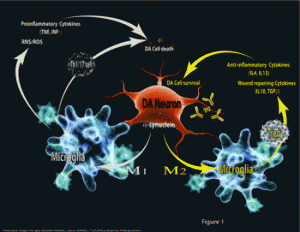 In the demyelination of multiple sclerosis, innate immune responses include microglia and other macrophages. When re-myelination occurs, microglia switch from the M1 pro-inflammatory state to the M2 anti-inflammatory state. Oligodendrytes are stimulated to make more myelin by M2 microglia and impaired if there aren’t enough M2.
Are Microglia the Most Intelligent Brain Cells
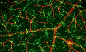 The remarkable microglia are involved in scavenging, phagocytosis, release of toxins and complex signals; they present antigens, prune synapses, control stem cells and brain circuitry, fight microbes and cancer, repair trauma, and respond to autoimmune diseases. Microglia have a vast number of receptors and signals that stay in constant communication with astrocytes, neurons, and all the immune cells.
Like the T cell, how can we not think that microglia are very intelligent cells?