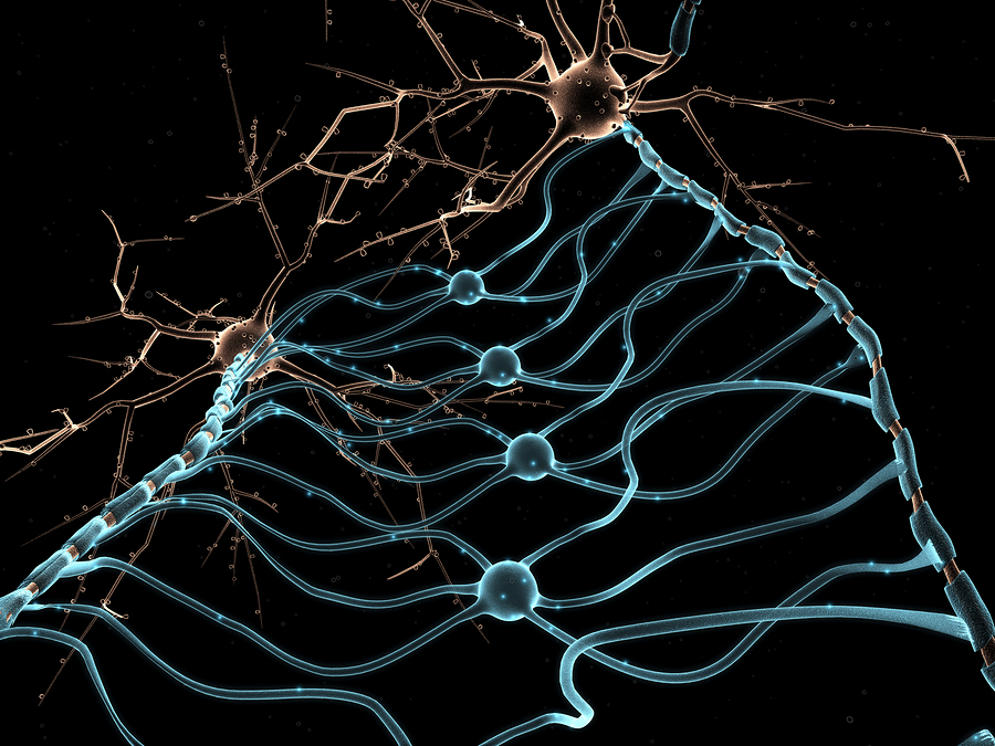
It is unrealistic to consider mapping the brain without most of the critical brain cells. The three glia—astrocytes, microglia, and oligodendrocytes—regulate all aspects of neuronal signaling networks. Many neuroscientists have focused only on the structure of neuronal connections and synapses. In fact one of the outmoded “dogmas” is that each neuron sends a signal to the next neuron, which along with other signals, is computed like a computer chip. This concept is hopelessly simplistic. There are thousands of factors in the neuronal network. And there are many other ways living individual neurons communicate—brain waves, electrical potential, cytonemes, and exosomes, among others.

Almost all of the current research incentives are for studying the details of the neuron electrical neurotransmitter network; and few to describe the three glial cells’ function. Brain mapping and “connectome” studies need to include all glial cells. Complications of the neuronal connection theory are presented in the post The Limits of Neuroscience; it includes the many other ways that brain regions communicate and the limitations of imaging. Recently, a new problem is described in a study of people lacking a corpus callosum. Despite this dramatic anatomical difference, the brain finds ways around the problem by communicating through other existing circuits.
The fact is that glial cells are absolutely critical in every aspect of neuronal function. The collaboration of glia and neurons occurs at many different scales—in large whole brain circuits, small local circuits and individual neurons and synapses.
Glia cells are very different from each other and from neurons, but they all share neurotransmitters, cytokines and neuro-modulation molecules. Glia provide growth factors like BDNF, Ephrin, TGFβ and TNFα, nutrients, metabolic regulation and ionic concentrations that determine the neuron electrical signals and blood flow. Glia determine the structure of neuronal connections, provide guidance for axons and dendrites and create physical barriers that provide direction for the structure.
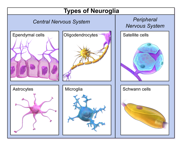 Astrocytes help create, mature, maintain and prune synapses and create factors that stimulate the production of new neurons. Microglia, also, prune axons and synapses, stimulate brain stem cells, communicate with all immune cells, along with a vast amount of other functions. Oligodendrocyte stem cells have the greatest amount of cell production in the entire brain and these are involved in creating the long-range and local structure of circuits while providing the myelin. Myelin is, in fact, extremely variable and able to produce molecules that inhibit axons and synapses in the white matter regions of the brain. The oligodendrocytes, also, provide regulation of the electrical signal in myelinated neurons.
All three glia cells stay in constant communication in many different ways. There is no brain mapping without glia.
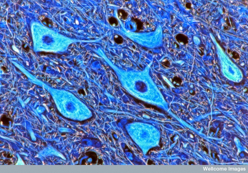
Astrocytes

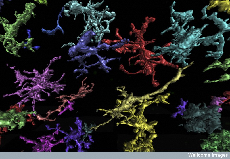
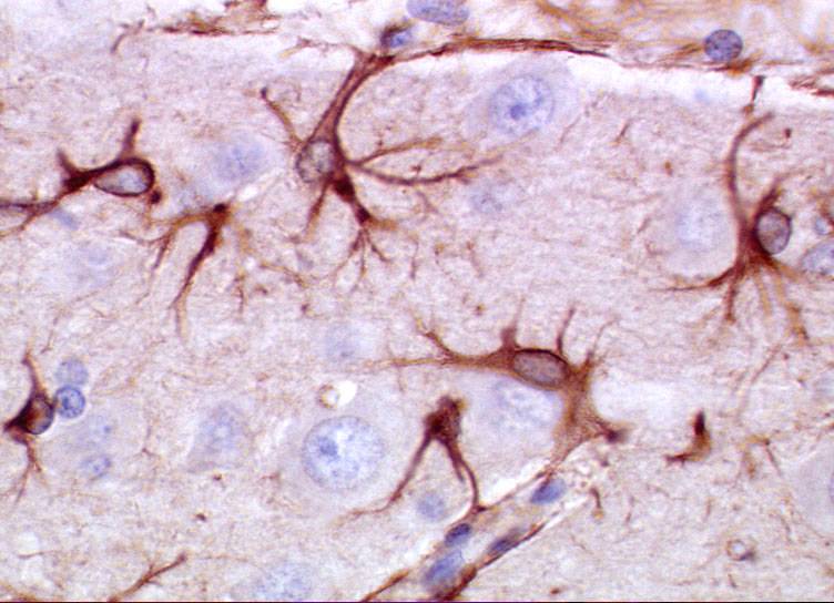
A previous post described the extremely complex vital support that astrocytes give neuronal networks and the entire brain. In fact, every aspect of the neuron’s function is totally dependent on astrocytes.
- Special astrocytes, radial glia, produce stem cells that produce neurons.
- More than half the brain is astrocytes.
- They maintain balance of critical ions, such as taking up potassium during action potentials.
- They pick up neurotransmitters like glutamate.
- They store the only energy in the brain in the form of glycogen, and determine the amount of glucose entering the brain through their connections with synapses and blood vessels.
- Astrocytes form a non-overlapping web structure that controls all blood flow in the brain.
- Neurons can have 100,000 synapses, but astrocytes connect thousands of different neurons at the same time in a much greater network.
- Astrocytes provide nutrients and digest waste for neurons.
- Astrocytes regulate synapse formation, maturation, maintenance, pruning and neuroplasticity through secreted signals and direct contact.
- Great complexity has been found in calcium signaling in multiple small compartments of the astrocytes in the many processes.
- Astrocytes use both electric gap junctions (see post on electrical synapses) and neurotransmitters.
- Oligodendrocytes have gap junctions with astrocytes.
Oligodendrocytes
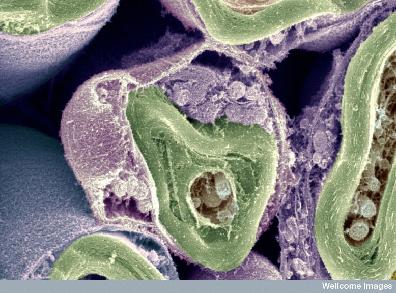
Oligodendrocytes have been thought to provide insulation for axons to propagate faster and not much else. Now, it is found that they, also, participate, along with astrocytes and microglia in very intelligent functions that are absolutely essential to neuronal function. Oligodendrocytes make important decisions about the types of signals that will propagate in a region. One of the new critical factors is the myelin code—the various different patterns of using myelin.
Myelin deposition is not a passive simple process. The structures of myelin, the number of layers of myelin, the length of the axon’s initial segment, the different lengths of the myelin in each part of the neuron, the size of nodes, and the large areas of un-myelinated axons, are all critical to determine the function of the neuronal network.
Myelin is not a simple insulator. It organizes the neuronal network and the types of information transfer. It allows areas for critical sideways local communication between neurons and immune cells. It determines the speed and flow of circuits. In fact, myelin forms a new critical code in the brain.
- Myelin patterns are different in each brain region, especially each of the six layers of the cortex.
- Un-myelinated axons are crucial of newly discovered types of communication between the naked axons and adjacent local immune and nervous system cells.
- Both myelin, interspersed patterns of myelin, and lack of myelin are essential for specific types of information transfer.
- In cortex layers II and III, there are different patterns of very long intermittent stretches without any myelin.
 The critical initial segment determines type and strength of the signal that is different in each region.
- The initial segment determines whether the spike can go backwards or not.
- Backward transmission determines aspects of neuroplasticity, such as memory consolidation in the hippocampus during deep sleep and quiet reflection when awake.
- Myelin plays a part in determining where synapses will occur.
- Many different regions of the brain have different amounts and distribution of myelin.
- Oligodendrocytes determine specific shape of the electrical spike and the amount of neurotransmitter release.
- Specific proteins in the myelin sheath stop the budding of an axons.
- Oligodendrocytes and its myelin determines the structure of the circuit.
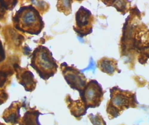
Microglia
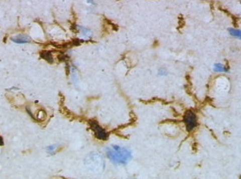 Microglia are perhaps the most intelligent of all individual cells and are completely crucial for the function of neurons. Individual cells each bridge many entirely different vital functions—scavenging, phagocytosis, release of toxins and complex signals, presenting antigens, pruning synapses, controlling stem cells, determining brain circuitry, fighting microbes and cancer, repairing trauma, and responding to autoimmune diseases. Microglia are the resident immune cells in the brain. They help establish neuronal networks in the fetus. They produce signals that nourish and stimulate neuronal growth and axon migration.
Microglia produce a vast number of receptors and use a large amount of different signals. These signals maintain constant communication with astrocytes, neurons, and all the immune cells. Microglia have many different shapes for these different functions.
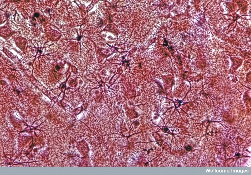
Each microglia cell maintains a very specific territory and travels independently circling the region. When they come to the edge of their zone they continue in their circular travels. During the travel they have arms that tap on every structure including neuronal axons, dendrites, synapses and astrocytes. By touching these structures they learn about what is occurring and can determine function that is not optimal. They know when they have to prune structures, like they known how to chase cancer and microbes. Their constant surveillance of the brain protects against any microbe invaders, demyelination, trauma and cancerous or defective cells.
 But, this is just the beginning of their amazing functions. They communicate using complex wireless signaling to neurons, astrocytes, oligodendrocytes and many different immune cells, determining how many brains cells are needed and when to eliminate a synapse—by eating the defective or unused synapses. They determine the rate of production of new cells and stimulate stem cells to provide this. In the fetus, they determine the amount of cells needed and eat the excess stem cells.
Microglia function is more complex than even T cells, but have many of the same talents. T cells were described in another post as masters of wireless signaling in the brain communicating to brain cells about when the “sick feeling” should occur and boosting cognitive ability before. T cells, also, travel through the entire body, including the cerebral spinal fluid. They chase invaders and communicate to other cells about many functions. They can rapidly change gears when they sense a problem and can suddenly produce large amounts of other cells. Microglia does all of this in the brain.
Glia Affect Large Brain Circuits
 Since neurons appear to sum inputs that arrive around the same moment, the time that a signal travels in a large network is an important factor. The conduction time of signals that travel through multiple regions is slowed considerably (hundreds of milliseconds) by several synaptic events of coding, integrating multiple signals at dendrites and the mechanisms of neuroplasticity. The actual conduction speeds are quite variable from less than one millisecond to hundreds of milliseconds. Glia regulate and modulate various links in a chain where complex coordination of conduction delays maintain the proper communication in long multistep circuits. Myelin increases the speed of the signal.
The production of myelin in different regions has been correlated with developmental milestones. Myelin in the frontal cortex occurs in young adults as they develop decision-making. But, in fact more subtle characteristics are constantly altered with experience, notably the new myelin code, the thickness of the myelin and the spacing of the nodes. Neuroplasticity is regulated in extremely complex ways. Now, it is shown that myelin patterns are, also, regulated with very complex signaling. As activity influences neuronal connections, myelination is influenced by signals from axons, membrane based proteins, and astrocytes regulating the ions producing electric signals.
Previous posts have described how neurotransmitters and cytokines are released along un myelinated axons in local regions. These are both in vesicles and freely secreted into the extracellular space. Adenosine triphosphate and tetrodotoxin (TTX) inhibits myelin and glutamate and leukemia inhibitory factor (LIF – from astrocytes) stimulates more myelin in neuron axons that are active.
 Another mechanism oligodendrocytes use to slow conduction time is by altering the axon, not the synapse. Glutamate and potassium channels create a depolarization in the oligodendrocytes that slows travel time. Other axons that do not have myelin are influenced by calcium from the astrocytes. This alters the shape of the electrical wave broadening it and altering the speed. This mechanism alters the axons not the synapses. A special astrocyte is associated with the myelination node of Ranvier and it alters the speed through potassium.
Glia alter the conduction time of neuronal action potentials by changing characteristics of the membrane potential and the timing of oscillations. Oscillations connect the actions of local and far away neurons together and influence the timing of the arrival of the various spikes at synapses.
Also, the great complexity of many overlapping brain waves is just being discovered. Astrocytes have a great affect on the membranes of the neurons have this has great influence on oscillations. Oligodendrocytes also influence the oscillations because of the long myelinated fibers actions.
The complex actions of the multiple glial cells affect the timing of signals not just in the range of milliseconds, but over hours and days.
Glia Affect Local Regional Circuits
Astrocytes are critical for all local synaptic functioning as described in a previous post. The local calcium signaling is described as extremely complex in another post including specific communications with small regions of axons, dendrites and the neuronal cell bodies. Astrocytes greatly influence whether neurons provide excitatory or inhibitory signals, such as regional inhibition during phases of sleep. They, also, regulate the potassium in the extra cellular space affecting the transmission of the action potential. They regulate every aspect of neuroplasticity changes.
Oscillations are utilized in local circuits and gamma waves (25 to 80 H) are highly connected with cognitive and motor functions. Astrocyte neurotransmitters from vesicles are critical for local gamma communication of this fast type. Future research must identify the astrocyte network and its production of these brain waves.
Individual astrocytes have very specific roles in small local circuits in multiple different ways. They use a variety of neurotransmitters and remove neuronal neurotransmitters in the synapses. They appear to have opposite roles allowing for regulation and homeostasis. Astrocytes release molecules into the space between the cells and each has a different effect. Adenosine decreases the neuron’s activity and glutamate increases it. This information is just now being discovered.
Astrocytes behave differently in different regions. In one hippocampus region decreased calcium is caused by particular neurotransmitters, like glutamate, but in others they respond differently. Some actions respond quickly; others need longer timed behavior of the neuron to respond. In some regions they respond through GABAB, various glutamate subtypes and TRPA1 channels. There are many other ways that different astrocytes respond individually.
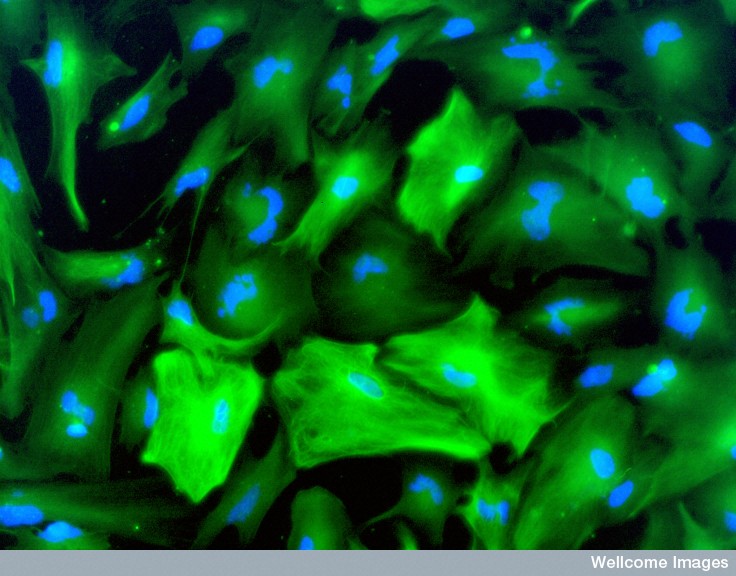
Astrocytes in Local Circuits
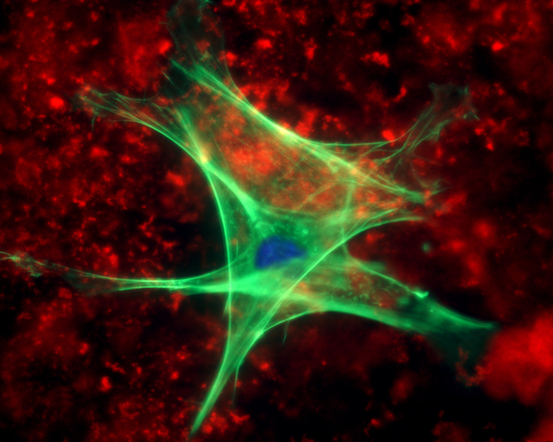
Human astrocytes are vastly more complex than rodent astrocytes often used in research. They, uniquely, contribute to human level cognition. Human astrocytes are 20 times larger and can coordinate a vast amount of synapses—up to millions. Like microglia they each have a territory that is not overlapping in the form of tiles. Since each is unique and integrates a large amount of synapses, they provide another unique network structure. They regulate the strength of the activity in this entire region. When human astrocytes replace mouse astrocytes, there was more neuroplasticity and better cognition.
Previous posts described how astrocytes regulate the creation, maintenance and pruning of synapses. They greatly influence the circuits and the connection network. But, it is not often appreciated that individual astrocytes in different regions behave uniquely. Where does this direction come from? Specific genes in different regions manufacture unique proteins. Therefore, these specific genes are somehow triggered in the region. Specific motor regions, for example, have unique astrocytes that affect the formation of circuits involving sensory data and motor responses. It is not clear how the astrocytes and the neurons can work so closely together to affect the meaning content of their circuits.
 Astrocytes regulate neuroplasticity and particularly dendrite spines. Astrocytes have many feelers in all areas of the synapse that can only be seen with the best electron microscopes. Actin that forms in these processes is highly mobile and responsive to calcium signaling affecting the complex tasks of regulating the synapse, the local circuit, the distance circuit and the meaning in the neuronal signal. These very small finger-like processes are more mobile when neuroplasticity is occurring. They can stabilize a specific dendritic spine to allow particular memory functions.
Astrocytes direct blood vessels in response to the needs of the cells in the local region directed by mental activity (this is what MRIs measure). The astrocyte feet on the blood vessels respond to the local amount of oxygen and activity to either open or close the vessels. They, also, send energy particles and ions back and forth between the neurons and the blood vessels. This provides support for the neuron’s metabolism. The effects range from small local changes to large region wide or brain wide changes. The astrocyte network can draw food and nutrients from far away in the network and use it for a local purpose. This becomes particularly important during a crisis, stress or seizure. If this process is interrupted then significant cognitive problems arise.
 A previous post showed how astrocytes are not monolithic and instead have many different ways that they signal with calcium. The calcium alterations can be global in the entire cell or can occur in one small finger-like region related to a single synapse. The large astrocyte has organelles that respond to a local synapse. The many processes protruding from the astrocyte have different calcium signaling when near specific individual synapses and participate in the neuroplasticity at that synapse. They are as responsive as neurons to the particular activity occurring at the synapse.
There are many important remaining questions. An important question is that of glia contribution to the electrical field potentials and synchronous oscillations. How is the enormous amount of dividing oligodendrocyte stem cells regulated? How do astrocytes and microglia keep in contact.
Imaging Glia
It is very difficult to image individual microglia. New techniques are emerging for astrocytes and oligodendrocytes.
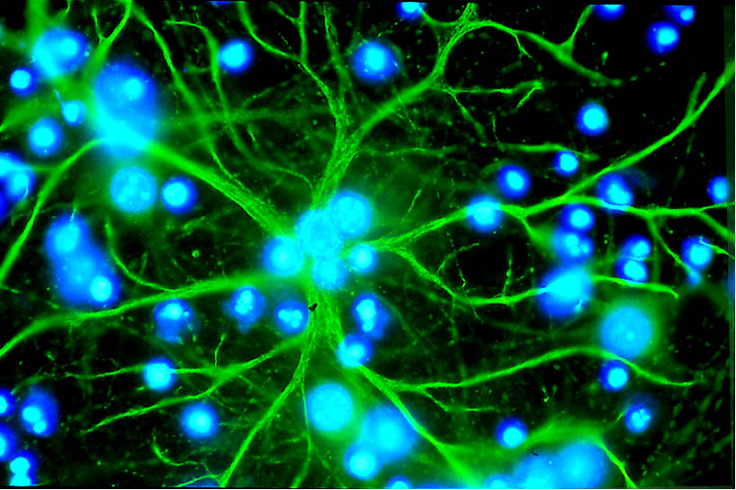
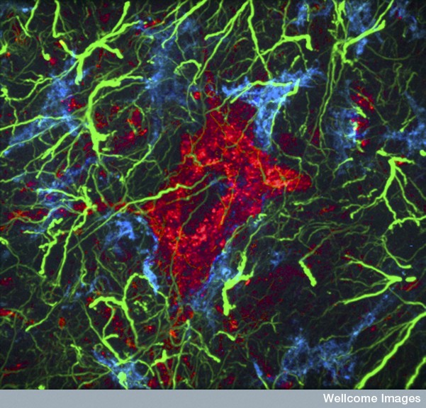
Imaging Astrocytes: Imaging calcium with fluorescent molecules that change with binding of Ca2+ ions have allowed observation of the interaction of glia and neurons. But these cells use many different forms of signaling that currently are not being directly imaged. Neurons operate with action potentials and can be measured using electrical membrane properties. Glia use a variety of receptors, channels and modulating molecules, which aren’t easily studied in the specific ways they are used in time and space. Current photo stimulation techniques are inadequate for real life applications. Another complication is that the same neurotransmitters are used by all the glia and neurons making it unclear which is using which. Most of the time glia functions are determined by studying the effects on the neurons.
Astrocytes use signaling among other astrocytes to limit their individual territories. It is not known how astrocytes compute information. It appears to be related to complex metabolic factors, not just calcium.
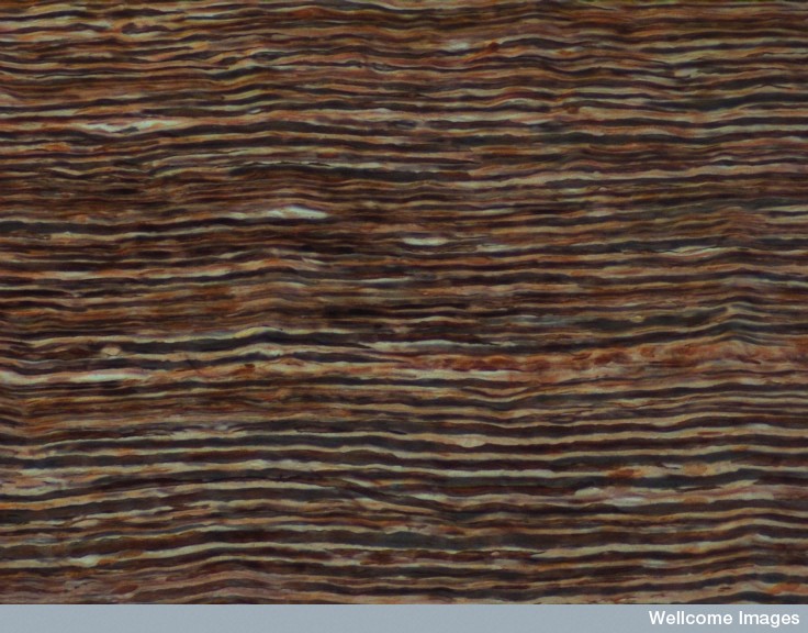
Imaging Oligodendrocytes: New high capacity diffusion tensor MRIs show that there are structural changes in the axons of the white matter, not just the cell bodies of the grey matter. It is not yet clear what is occurring, but there is evidence that myelin, oligodendrocytes and astrocytes are involved. While very unique myelin codes have been described in the cortex, most regions have not been analyzed. Neuron-centric bias has limited understanding of the real complexity of myelin. It is very difficult to study oligodendrocyte activity, since it involves microscopic changes of the details of the myelin over the scope of large whole brain circuits. When this vast amount of data is analyzed new discoveries are significant, such as cortical layers of II/III, IV and V with different myelin codes.
Another complication is that the electrical signals along the axon can go backwards as well as forwards and this is affected by the specific myelin code. Specific versions of the initial segment are particularly important and variable. (see post on Complexity in the Myelin Code). Myelin determines where synapses can occur and not occur. New imaging devices need to be developed to more accurately identify myelin
No Brain Mapping Without Glia
It is impossible to consider understanding brain function without the three glial cells. To think that just mapping the neuronal connections will describe mental function is absurd. While most money is going into studies of neurons, there is a vast amount of work to be done on the three glial cells.
Astrocytes provide nourishment, control blood flow to the neurons and participate in the information transfer with its own system; astrocytes, also, establish a protective structure for the neuron. The astrocyte network is as vast as the neuronal circuits, if not more, and has its own type of signaling that is just being discovered.
Microglia travel throughout the brain participating in surveillance, stimulation, cleanup, and maintenance tasks with wireless communication to and from all other cells. They regulate stem cells in the fetus and the adult brain. Individual intelligent microglia patrol the brain like T cells and respond in complex ways to many situations.
Intelligent oligodendrocytes are not passive insulators, but provide a wide variety of different patterns of myelin, both in location, size and shape. They help determine the conduction rates of neuron circuits, such that relevant information can arrive at appropriate times. They are critical to create the structure of neuronal circuits.
Mental events are correlated with all of these glial systems and the neuronal circuits at the same time. Mental events stimulate neuroplasticity, which occurs instantly and simultaneously in many different parts of large brain circuits using many different mechanisms, determined by the cross talk of these four interacting systems. Mental events are related to the communication of individual intelligent cells performing vastly complex functions together. Where is the direction for all of this?