 There is increasing media attention to conclusions from neuroscience research, many of which affect society. Unfortunately, often these conclusions are not warranted from the current level of knowledge. All types of theories about the brain, the mind, free will, ethics, and morality are circulated as facts because one of the 500,000 neuroscientists have espoused unproven and speculative viewpoints. Several books recently have pointed out that results from fMRI are, especially, being over interpreted. This post will address some of the limits of current neuroscience.
One important problem with almost all current fMRI studies was recently noted. It concerns a critical assumption in the design of most experiments that is not correct.
Each study has a group that performs a task, and another control group that it is compared to. The assumption in the experiment is that the group performing the task activates a specific part of the brain, which is an addition to the control group’s brain activity. This is called “cognitive subtraction.” It assumes that the control group’s brain state participates in some part of the task group’s brain state. An example could be the resting state during day dreaming (called the default mode network) compared with the brain state during a specific action. The new brain state is compared with the resting state and one is subtracted from the other. The new activity observed is assumed to be the brain regions of the action.
However, recent studies show that, in fact, many tasks use cognitive “addition” where parts of the brain do less when the experimental task is done. That is, it cannot be assumed that none of the resting state, some part of the resting state, or all of the resting state are used in the task. This simple assumption might invalidate many current studies and conclusions.
But, this is just one of many problems.
What is Subjective Experience?
 A fundamental problem in current neuroscience is that there is no known center of subjective experience in the brain, and there isn’t any understanding of what it might be. There has been no evidence, except is very special cases, that the changes observed in imaging devices actually are related to subjective experience. Most scientists simply ignore the question of what the mind might be because the answer seems impossible at this time with current molecular theories. Most just assume that somehow, the networks of electrical impulses create the subjective experience of mind.
Much of what we think we know about mind comes from analyzing damaged areas in the brain and what this does to cognitive capacities. While these damaged areas are clearly necessary for mind to work there is no indication that they are mind. These areas might be a molecular device that the mind uses and directs.
Recently, a leading neuroscientist noted in Nature:
“The general public might think that this goal has already been achieved; when they read that a behavior is associated with some part of the brain, they take that statement as an explanation. But most neuroscientists would agree that, with a few notable exceptions, the relationship between neural circuits and behavior has yet to be established.“
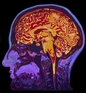
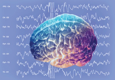
fMRI Imaging
Since so many results today depend upon fMRI, several other problems need to be discussed.
There is little direct evidence that fMRI as currently used measures neuronal activity.
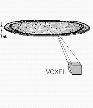 What fMRI measures is the blood flow in a region of neurons. Each dot of light on the fMRI, the voxel (the pixel of fMRI screens), captures blood flow in the region of approximately 80,000 neurons and more than 4 million synapses. This dot of light on the screen is the average measurement of activity over one second in this region. However, neuronal signaling occurs a thousand times faster, in the realm of milliseconds.
 Astrocytes, not neurons, are the cells that determine blood flow by having feet on each vessel and opening and closing them to provide local neurons with blood (see post on astrocytes).
By the time the blood flow is measured in the region of the 80,000 neurons, signaling in milliseconds has occurred in large circuits around the entire brain, none of which is picked up at all. There is no way, really, to say that the mental event is correlated with this voxel (blood flow in the region of 80,000 neurons), instead of one of the thousands of small events that cannot be measured. Also, it is not known how the astrocytes changing blood flow really reflects the action of neurons.
What are Valid Studies?
 Just to see how relevant most research is, two recent studies did something very unusual. Instead of the small number of subjects usually used in the neuroscience studies that everyone reads about—fifty subjects being a very large number for an ordinary study and usually only a few repetitions of the action—these studies used more than a thousand people in one, and 500 repetitions of the action in the other.
 Almost all of the events that other studies would have correlated with one specific brain region were found to be incorrect. The routine studies are based on a small number of subjects or repetitions and show the “strongest” regions. These much more detailed studies showed that events, instead, occurred in many very small regions all over the brain, which were not picked up by the conventional studies. Because there were so many small events, none of them could be correlated with the subjective experience.
Also, conventional studies chose a specific moment for the effect to be measured and then chose the largest effect at that second. In fact, the larger studies noted many small events in sequence before and after that supposed “moment” of the event in much smaller time scale. The vast majority of reported studies simply cannot pick up the complexity.
Both of these studies concluded that current devices are not adequate.
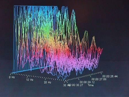
In another study when attention was changed, the entire brain changed, not one region. The only way to understand the total brain activity was to know what the person was thinking about.
 Another study looked at the brain waves and oscillations of the entire brain during activities such as tapping a rhythm with fingers. The study found rolling activity over the entire brain for activities that had previously been thought to be located in one motor region. This rolling wave was not random but had a specific pattern for each different activity. In fact, repeating an action showed different patterns since there was learning from the first tap to the next – conscious activity is eventually trained in the habit memory regions to become an unconscious behavior.
These are not just my conclusions. Many of the leading neuroscientists are alarmed at the false conclusions in the popular press, and the over reach of scientists in the popular media. These oft repeated firm conclusions give a very inaccurate picture of what is really known.
Recently, in Nature Reviews Neuroscience, it was noted that most neuroscientific studies have too small a number of subjects to be certain of the results. It notes:
“that the average statistical power of studies in the neurosciences is very low. The consequences of this include overestimates of effect size and low reproducibility of results.”
A similar recent sentiment was given in Scientific American, “An Epidemic of False Claims” where it is stated:
“False positives and exaggerated results in peer-reviewed scientific studies have reached epidemic proportions in recent years. The problem is rampant in economics, the social sciences and even the natural sciences, but it is particularly egregious in biomedicine.”
New Types of fMRI
There is a new type of fMRI being developed using different “diffusion” techniques that measures molecules that diffuse—mainly water in biological tissues. In this new type of software analyzing the fMRI data, the computer algorithm gives a best guess of water diffusion rates at the spot that represents the voxel. With this technique it may be possible to measure changes in neuron’s membranes, the water inside the cell and the water outside the neuron’s membrane. This picture might be more accurate, but it remains to be seen exactly what will be measured.
Complexity of Brain
 There are many other difficulties in understanding the brain. Two previous posts detailed many technical problems with the search for the neural code and the map of the connections. (see post Neural code, and the Connectome) These technical issues are problems that the big brain projects in the U.S. and Europe will need to confront. Please refer to these posts for more details.
The following is a list of important issues raised in previous posts that greatly limit current neuroscience and affect our ability to make the types of statements that are frequently made in the media.
- Changing moment-by-moment neuroplasticity with new and pruned synapses each day.
- New synapses placed in new spatial relationships on the dendrite
- Large numbers of different types of neurons in different brain regions
- Extremely complex extracellular matrix
- Postsynaptic densities that are different in different regions and each include thousands of large complex interlocking proteins.
- Individual neurons participate in different neuron subgroup circuits for different tasks.
- Small RNA molecules are transported in exoxome sacs from neurons and picked up, like a virus, by other neurons, even quite far away where they are able to alter function.
- Information transfer between neurons is both hard wired and by wireless synchronous oscillations.
- Wide range of neuroplasticity mechanisms simultaneously in large brain wide circuits (see post on Neuroplasticity)
- Subjective mental experience relates to 12 orders of magnitude of size, from quantum, molecules, cells, organs, brain, society (see post Life of a Thought in the Brain.)
 Multisensory: Previously it was thought that there are specific sensory regions for sight, sound, etc., which are then relayed to other associational regions. All of the algorithms have made this assumption. Recent research shows that, in fact, most neurons are multi sensory or multi modal—that is, they are connected to many different senses or modes at the same time. Also, the modular theory of the brain assumed that there were many local hubs with vast interconnectivity but limited connections to far away hubs. Now, it has been shown that most hubs have both vast local and distant communications, making theories of mind through neuronal connections much more complex. This also makes a search for a center of subjective experience much more difficult.
 Electricity in the Brain: Electricity in the brain occurs in many ways (see post). In addition to the well known axon spike, there are synchronous oscillations of large numbers of neurons; electrical gap synapses; local field potentials in the extracellular space; ephaptic communications from the sides of un myelinated axons to immune cells; calcium waves; and the astrocyte communication network that is larger and as complex as the neuronal network and determines all aspects of the neuronal synapse as well as fMRI information.
Memory: Memory is not a single process but exists in broad circuits around the brain. Very recently, it was found that in a mouse, long-term memory appeared to be in the cortex, not in the hippocampus, where it had always appeared to be. This study disabled critical NMDA glutamate receptors and showed that disabling them in the hippocampus did not alter the long-term memory but disabling them in the cortex did.
There is really very little known about how a single memory is formed. A recent study showed that two specific regions send memory information by synchronous neuronal oscillations. The study focused on time and place, two key elements of an explicit memory of a scene – where and when it happened. Remarkably, brainwaves showed two different frequencies from the involved regions, one for place and one for time.
Laboratory Studies of Animals
Another big set of problems for current neuroscience is, rarely, ever discussed.
Research, usually, depends upon having a way to study a living brain. The only time that live human brains can be used is with very crude devices like fMRI, EEG, or, occasionally, with neurosurgical patients who have their brain opened for surgical procedures and agree to allow electrode probes of the effects of stimulation of different brain centers.
In all other cases, neuroscientists study animal brains and try to develop a model of a situation that the human brain faces. This research often mutates genes in cells of different animals (flies, worms, mice, etc.) that seems relevant to a human disease. This process then extrapolates from the animal to the human.
 But, a major problem is the inherent difference in the research results when studying animals in the captivity of a laboratory compared to animals in their natural surroundings. While it is obvious that larger, highly intelligent animals such as apes, elephants and dolphins, behave differently in captivity, it has only recently been found that this applies to many smaller animals as well. See post for details of mental illness of captive animals and the ethical questions this raises.
Also, recent research shows that even very small animal brains demonstrate high intelligence, advanced social behavior, and unusual capacities that human’s don’t have. These capacities occur in brain regions that are very different from the human. So, simple comparison with activity in human brains is speculative. (See post, Animal Intelligence Update).
 Suprisingly, recent research has shown that many of the favorite small animals that are used for neuroscience research—zebrafish, flies, worms, mice and rats—also have dramatic differences in results when the research is done in the laboratory versus natural settings.
Housing conditions of zebrafish can have a major influence on scientific results. Recent research of mice in laboratories showed that providing a natural environment had an impact on the results of many unsuccessful trials  of medication. Also, with flies, important differences are seen between their actions in laboratory settings and in the wild. In one lab study flies followed circadian clocks, but, in nature they followed the natural light. A conclusion of the scientist was that not only are natural surroundings necessary for correct research results, but also for identifying what would be best for the animal.
Studying animals in natural settings is very difficult. Without being able to understand humans’ inner world, extrapolation to other species is very speculative.
The Limits of Current Neuroscience
For the many reasons outlined, current neuroscience, while exciting, is over reaching in many of its conclusions. There are many popular writers of neuroscience (both scientists and journalists) who have made far ranging conclusions about the mind, ethics, morality and philosophy supposedly proven by neuroscience. Most of these are unwarranted by the evidence.
 As can be seen, there has been no progress in finding any region or structure of the brain that explains subjective experience. In fact, the evidence does not support that there is going to be a molecular explanation of mind at all. Much of the data on this website in neuroscience, animal intelligence, plant intelligence, and microbe intelligence, including recent molecular biology and genetics, points in the direction of mind being a property of nature, such as spin or charge in small particles. Perhaps, by adopting the new viewpoint that mind exists throughout nature and interacts with various molecules, cells and brains, the current deluge of research can be better understood.