
Both the wired neuronal brain and the wireless brain of the immune system perceive stress. The fact that psychological, social and physical stress all trigger responses from both the immune system and the brain, shows that immune and brain systems cannot be separated. Significantly, stress also triggers multiple hormones. Dealing with stress can be a lot for anyone to handle, especially if you are not sure how to manage it. When one stress event occurs, the brain adapts with neuroplasticity responses altering the way other future stressors are handled.
Recent research shows that the type of neuroplasticity that occurs in the hypothalamus from stress is unique and starts with the first event. There are complex ways that future stressors can cause of a variety of outcomes. This unique type of neuroplasticity is based on time windows with different types of effects immediately after, several hours later and days later.

Stress alters synapses to neurons in the hypothalamus that produce stress hormones and these changes limit future ability to adapt because of neuroplasticity. The particular neuroplasticity stress induces is extremely complex and just being worked out. It involves changes in glutamate synapses, as well as bi directional alterations in the signaling of cannabinoid and GABA systems.
These embedded forms of neuroplasticity are called metaplasticity– altering the ability to be plastic in the future. In this system, a unique type of metaplasticity allows future changes at particular times after the first incident and is called kairoplasticity (for the opportune time.)
Responses to Stress

Stress is the body’s reaction to a threat, either real or imagined. It requires immediate attention and change. The body responds with fear, anxiety, flight and flight and stimulation of hormones. These reactions are important in decision making of all types. Extreme stress is highly connected with depression.
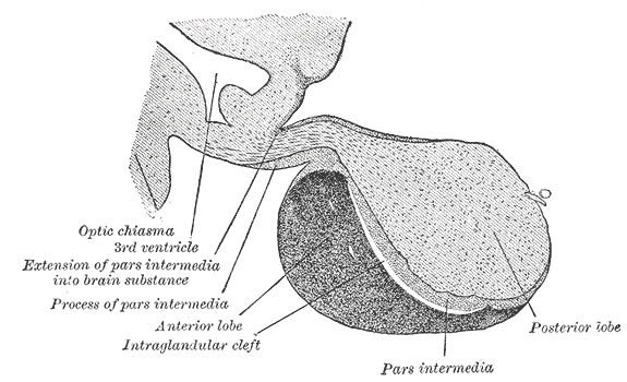
After a stress, there are adaptations and alterations that prepare for the next stressor. Well known regions of the brain that respond to stress are called the HPA and include the pituitary, adrenals and hypothalamus (specifically, the paraventricular nucleus of the hypothalamus or PVN). Many other learning, memory and emotional centers are involved, as well.
Neurons in the PVN respond to stress by secreting a hormone called CRH (corticotropin-releasing hormone) after integrating information about the stress from many other parts of the brain. Other nearby cells secrete other hormones, as well. All of these neurons are known as PNCs or parvocellular neuroendrocrine cells. Various neurons send their hormones in a special brain blood vessel called the hypophyseal portal circulation.
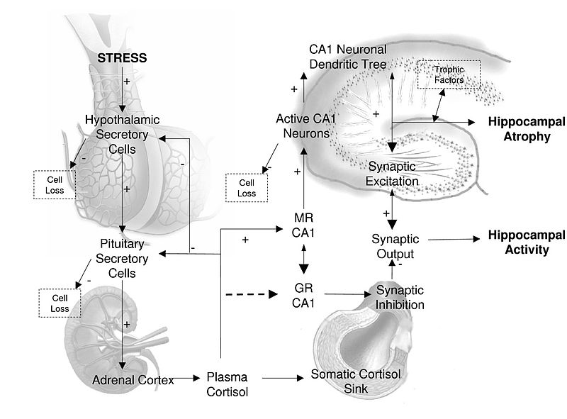
CRH triggers another hormone called ACTH (adrenocorticotropic hormone) from the pituitary that enters the general blood stream. This hormone travels to the adrenal cortex stimulating the release of corticosteroids, which have wide-ranging effects on metabolism throughout the body.
This complex brain mechanism to stimulate steroids allows elaborate feedback and control of very important powerful hormones. Complex signals from many parts of the brain (amygdala, hippocampus, cortex among others) are integrated by synapses on the PNCs, which control and modify the responses from the hypothalamic-pituitary-adrenal HPA axis. When this system is triggered, glutamate synapses and GABA synapses on the PNCs engage in elaborate neuroplasticity to respond to the stressors.
Very Brief Outline of Neuroplasticity
 This post will discuss the remarkable and unique type of neuroplasticity that occurs on critical neurons (PNCs) in the hypothalamus that are responsible for an immediate and long-term response to stress.
Previous posts have described the extremely active brain, which creates new neurons for memory and learning and builds and prunes a fantastic number of synapses every day and night. Research shows that neuroplasticity builds stronger circuits with particular meaning and responds instantly to mental events. What is remarkable about neuroplasticity is that it occurs in wide ranging circuits throughout the brain with many simultaneous different mechanisms. A list of some of these mechanisms is given here.
Types of neuroplastic changes that can occur simultaneously in synapses of large circuits:
- Changing myosin motors and actin structures
- New AMPA glutamate receptors
- Changes in complex post synaptic density (1000 interlocking proteins)
- Changes in surface molecules sticking from
- Calcium surge triggers new types of “memory proteins”
 Substitution of new matching neurotransmitters and receptors
- Alteration of signaling cascades from the membrane to the nucleus
- Changes in axon ion channels altering electrical signal
- Change in balance of inhibition and stimulation
- New micro RNA’s alter process
- Mitochondria change strength of the signal
- Climbing fibers in cerebellum with multiple alterations
- NMDA glutamate receptors substitute their subunits –
- Transport motors are substituted
- Actin and microtubules direct scaffolding structural changes
- Exosomes information from astrocytes to neurons with DNA and proteins
- Changes in extra cellular matrix
- Potassium channel changes
- Neurons causing inflammation in large synapses
Stress and Neuroplasticity
The effects of stress on the brain are extremely complex with many different kinds of neuroplasticity in different brain regions. Different effects occur for individuals (varied resilience) and from various factors of duration, intensity and environment. Stress responses occur in all animals and elaborate mechanisms have evolved to deal with it at all physical, emotional and social stress. These mechanisms involve changing genetic networks and epigenetic tags on DNA and histones. Even a brief exposure can have lasting effects.
There are different patterns in varied brain regions related to memory and learning that coincide with the changes in the hypothalamus that are described in this post. A very brief summary is:
Amygdala:
- An increase in neural activity
- Increased dendrites and spines
- Increased long term potentiation
- BDNF is increased
- More neuroplasticity
Hippocampus
- A decrease of neural activity
- Decrease of dendrites and spines
- Decreased long term potentiation
- BDNF decreased
- Less neuroplastiticy
Pre Frontal Cortex
- Decrease of dendrites and spines
- Decreased long term potentiation
- BDNF increase or decrease
- Messenger RNA increase or decrease
- Weakened connectivity
- Changes from reflective to reflexive
- Disruption making cognitive disorders worse
This post begins the discussion of stress related brain changes by describing unique neuroplasticity that occurs in the hypothalamus. Future posts will discuss neuroplasticity from stress in the prefrontal cortex, hippocampus and amygdala.
PNV Connections
 PNVs have many types of inputs, but mainly fast glutamate and GABA neurons. They, also, have synapses from other types of neurons with monoamine and peptide neurotransmitters. Monoamines include histamine, epinephrine, norepinephrine, dopamine, serotonin, and melatonin. Peptide neurotransmitters include somatostatin, substance P and opiods.
Neurons that synapse onto PVNs have many clear round vesicles for glutamate and GABA and as well a different type of vesicle called “dense core” with amines and peptides. It appears that the peptide and amine neurotransmitters are co transmitters with glutamate and GABA. There are, also, slow modulating neurons as well.
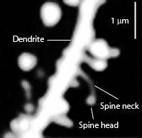
In most parts of the brain, asymmetrical glutamate synapses consist of up to 60 to 80% of the synapses. But, in PNV half are symmetric GABA inhibitory synapses (for comparison, the hippocampus has 3% and visual cortex 6%). Thirty percent of asymmetrical synapses use epinephrine and norepinephrine neurotransmitters as well. All of these synapses can be connected at dendrites, axons or cell bodies and are very close, intermingled and complex. The dendrite is simple and smooth with few spines, unlike other brain regions. In the amygdala there are ten times as many spines.
These special synapses are so close to each there is spillover of the neurotransmitters into nearby synapses, such that glutamate and GABA affect each other’s actions locally. This allows a very unusual communication between both.
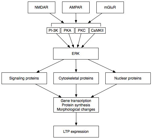
There are multiple kinds of glutamate receptors active (AMPA, kainite, NMDA and metobotropic) as well as GABA with various subunits (A, AR and BR). Glutamate seems to stimulate CRH. GABA maintains a baseline inhibition that can be released.
Stress Anatomy
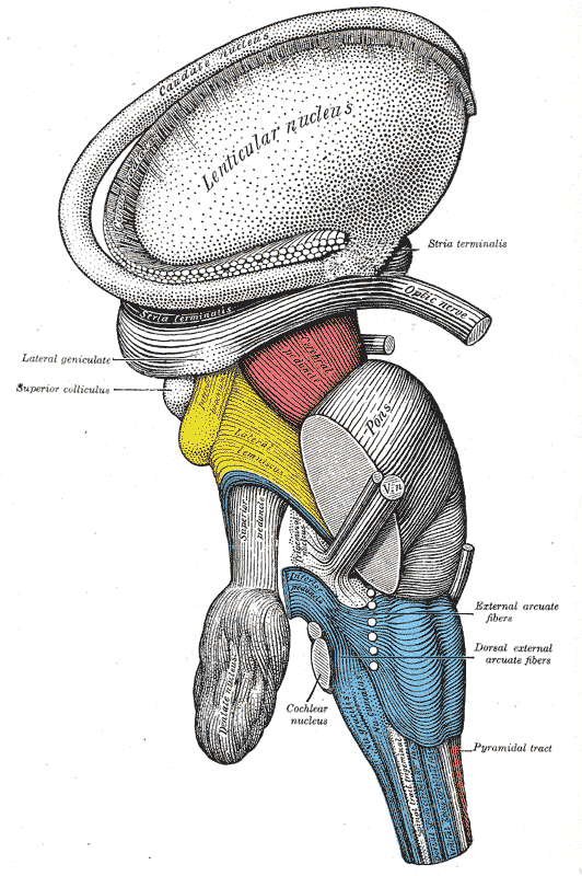 A circuit from multiple parts of the brainstem and parts of the hypothalamus sends information about bodily functions and states to the PVN. In fact, a large number of very complex circuits have been found that have input into decisions made at the PNV. Psychological and social information comes through many different circuits including limbic regions, hippocampus and pre frontal cortex. These connect to structures in the hypothalamus and then to the PVN. Circuits run through many deep brain structures from all over the brain and are finally integrated in the PVN in relation to stress.
One major source for synapses at the PVN is the BNST (posterior bed nucleus of the stria teminalis). The BNST is highly connected to the amygdala and other limbic structures. It is active in anxiety and threats and connects with the orbitofrontal cortex related to unfamiliar people. Other connections are brain stem tracts with inputs related to norepinephrine and epinephrine. The circuits are extremely complex and just now being discovered.
Glutamate Synapses in PVN
Glutamate neurons make many different subunits for receptors. Switching these subunits has been noted to produce particular forms of neuroplasticity. (See post on Neuroplasticity by Altering Receptor Subunits). In the PNV subunits are altered and up regulated after stress. Norepinephrine modulates these glutamate synapses regulating stimulation of HPA. During stress, it increases the glutamate activity. They increase ACTH and corticosteroids.
Brain cannabinoids are a vital part of the PVN synapses. (See posts on cannabinoids in the brain for a discussion of their vital role in many brain circuits.) They are released by the postsynaptic neuron and act retrograde back to the first neuron. They inhibit the release of glutamate and GABA slowing the HPA secretions. When PNCs are highly stimulated, more cannabinoids are manufactured to tone down the activity. The more corticosteroids produced, the more cannabinoids are produced to decrease the activity. This occurs through elaborate cascades and genetic networks.
After Stress
 One stress event has an effect on the glutamate synapse in the PNC. The ratio of two different receptors is altered with more AMPA and less NMDA. This occurs by a great decrease of the activity of the NMDA glutamate receptors. CRH local release causes a decrease in the NMDA receptors, either by the same neuron or from other cells. This decrease of NMDA stimulates an unusual form of neuroplasticity that is dependent on the type of activity that is present.
This new type of neuroplasticity is called metaplasticity and is stimulated by bursts of activity and then a short-term alteration in the synapse. The term plasticity refers to the alterations that occur at a synapse. Metaplasticity is a change in the ability of the synapse to have different kinds of plasticity in the future. It produces plasticity that is either an increase or decrease of activity for future mental events. This metaplasticity continues to affect future reactions to stress. It increases the responses to the next stressors. After just one stressor, this effect lasts for many days.
Metaplasticity includes a different way that glutamate vesicles are released. Before the change, one vesicle of glutamate is released at a time, and after the change multiple vesicles are released at once. Normally, the release of multiple vesicles is blocked by retrograde inhibition. CRH decreases the amount of calcium signaling in the NMDA, which blocks the inhibition allowing the multiple release of vesicles. This retrograde signal, surprisingly, is not from the cannabinoids.
There are several other responses to the stress that are independent of the multiple vesicle release at NMDARs. These, also, contribute to metaplasticity. Multiple stress events decrease signaling of cannabinoids and therefore stop the tonic inhibition. This decrease of the cannabinoid signaling is activated by steroid receptors. Norepinephrine and corticosteroids increase several of the glutamate receptors’ activity at the PNC. This change from stress alters the GABA synapses in a similar way to the CRH metaplasticity at glutamate receptors.
These three mechanisms create a completely different way that the synapses operate, altering future responses to stress.
Long Term Changes After Stress

Response to stress alters HPA activity, becoming overly responsive and sensitive to the next stressor. This appears to involve an increase in the glutamate synapses, but the mechanism is not totally understood. In animal experiments, multiple stressors in mice increase the number of synapses and dendrites at the neurons that secrete CRH. The number of connections to the PNCs is doubled in both glutamate and GABA. Continued multiple stressors in young life completely alter the activity currents of the synapses increasing reactivity.
Multiple stressors produce increased activity at baseline and more extreme responses. Another change occurs in the astrocytes with less transporter molecules for glutamate. The astrocytes are vital to the increased sensitivity in these HPA neurons.
The reduction of stress can, also, have effects on neuroplasticity, but this has not been well studied. It can lower the increased sensitivity, but only in certain windows of time. In one experiment with mice, increased environmental nurturing decreased stress response. The number of glutamate synapses was decreased. Early life activity appears to produce epigenetic alterations that last into adulthood and might be beneficial or detrimental in this regard.
GABA synapses are fifty percent of the activity and are inhibitory. Acute stress decreases GABA signals at PVNs. There are many factors involved in this and the exact mechanism is not clear. Transporters are decreased for reuptake and chloride channels are altered.
Different Effects at Different Time Windows
GABA synapses are, also altered with a unique type of neuroplasticity. What is unique about this type of metaplasticity is that it is correlated with timing after the first stressor. Without stress, GABA synapses at PVN do not have long lasting neuroplastic changes. But, after either physical, psychological or social stressors, neuroplasticity changes of several types occur—both long term potentiation and the opposite long term depression.
Whether potentiation or depression occurs is based on the timing—potentiation immediately after stress and depression 90 minutes later. These opposite directions of the neuroplasticity come from competition between corticosteroid effects (long term depression) and norepinephrine effects (immediate potentiation).
Beta-adrenergic receptors alter pathways stimulating specific glutamate receptors. This creates a time window and then other glutamate bursts cause the change. Corticosteroids stimulate receptors on the PNC cell body causing a cascade that produces glutamate signals that stimulate opiod peptide encephalin release. This inhibits GABA in retrograde manner.
With chronic stress, GABA synapses are altered by decreases of the tonic inhibition from cannabinoid retrograde signaling. Synapses change their architecture. More GABA synapses appear, especially at dendrites rather than the cell body. Strangely this new configuration actually has less GABA activity in a long-term manner, such it is thought that many of these new synapses are silent. Somehow, this alters the HPA output.
PNC Synapses Are Very Active
Critical PNC neurons have extremely active synapses that undergo rapid changes in response to psychological and physical stress to the organism. They are radically changed based on experience. These neuroplastic changes have dramatic effects on the HPA stimulation of stress hormones.
Alterations appear to be related to chloride channels and flow across the membrane of the neuron. The exact mechanisms are very complex and not understood. But, norepinephrine, a molecule associated with sympathetic fight or flight responses, stimulates overly sensitive HPA secretion. Corticosteroids from the adrenal slow down the HPA by feedback loops.
The neuroplasticity of critical PNCs is very complex with overlapping and opposing mechanisms. The results have extreme relevance to how one stress experience predisposes people to future stressors. It is relevant to resilience. The very first stressor sets in motion multiple opposing cascades that operate based on further experience.
It is not yet clear how the time windows operate, but there are different processes that occur just after the stress and later on. These windows are bidirectional in that the system can go in different directions. It is one of the first known brain regions where timing is a critical part of the effect of neuroplasticity. This fits with our experience that there are many different kinds of responses to one or more stress experiences. There are immediate effects, 90-minute effects and alterations that occur three days later.
Some evidence shows these processes can be reversed with less stress, possibly with relaxation and meditation techniques. There is evidence that the meaning of the event is critical to either increase the stress response or do the opposite and this is built into the synapse. But, not enough research has been done.
Unique Type of Neuroplasticity With Stress
 Everyone is aware of how critical stress is in our lives. It can stimulate positive accomplishment or long lasting damage. It can affect the immune system, physical organs, and cause depression and anxiety. The inseparable relation of the brain and the immune system is just being discovered. Psychological, social and physical stress immediately affects the brain, the immune system and many hormones. Mental events have immediate effects on all of these biological systems.
The brain is extremely active with daily alterations in synapses and new brain cells for memory and learning. Stress stimulates unique forms of neuroplasticity. One type alters the synapses so that they respond differently with new types of neuroplasticity in the future. Another type appears to work differently at various times—immediately after a stress event, hours later and several days later. These findings are consistent with our experience that stress can have many different effects.
Mental events trigger immediate responses, not only in the brain, but in immune cells of all types. These mental events trigger immediate and long lasting changes in brain circuits. Neuroplasticity changes at the PNCs are responsive to the meaning of the stressor. This demonstrates mind interacting with neurons, synapses and chemicals.