 When considering the intelligence of individual cells, certain things stand out. Cells know exactly where they are and how to travel to far away places through many kinds of terrain with many different techniques. Cells know how to signal back and forth wirelessly to many different types of cells using many different signals. Cells know how to become a particular type of cell and stay that way. Today’s post will discuss another important type of cellular intelligence—how do cells know what size they should be.
Cellular Intelligence
 There are many aspects to cellular intelligence that have been described in previous posts. Several of them refer to a cell understanding its relation to the external environment and world without GPS. These have been described in posts on how a cell knows where it should be and how it knows how to travel long distances over complex terrain to exact places.
It is remarkable that a cell can know exactly where it is in a large world of billions of cells. For this, cells need to make complex calculations and decisions based on its relation to other cells. Immune and blood cells need to know exactly where trauma or infection is located and how to get there. Neurons find very precise locations in the fetus and the adult brains. Growing cells in limbs and organs must know exactly how they fit into the shape and where the edge should stop.
Cells use multiple measurements and rules to calculate, but they also have to adjust for many sources of error in this process. Data comes from diffusible molecules of morphogenic fields, electrical gradients and networks of genes. They signal to other cells to validate or amplify this information. (See post on the Remarkable Language of Cells). Cells take measurements and then check with neighbors. Measuring a gradient is difficult with many accuracy problems. Cells use multiple feedback loops, timed measurements, gene regulation, signaling and self-organization with other cells. Each situation has many variables and cells become virtual mathematicians to synthesize the information.
To travel to far away places, leukocytes, T cells and other immune cells use thousands of different factors and many different modes of travel. They change their size for different terrain and situations. They signal back and forth for advice and information from blood cells, neurons, endothelial cells and immune cells; in each type of bodily organ the communication is different with a large number of cytokines and chemokines.
Leukocytes navigate the blood vessels by grabbing onto the lining with adhesion molecules, then rolling and tethering. They sneak through the cells into the tissue with many techniques. They, then, respond to positive and negative gradients of all kinds. Each cell can use multiple different kinds of movement and different shapes, triggered by genetic networks operating actin-myosin motors and scaffolding structures. Travelling cells can use their leading edge in different ways and can create swarms of large amount of cells in one particular spot. They navigate the extracellular matrix and other obstructions.
Cell Size
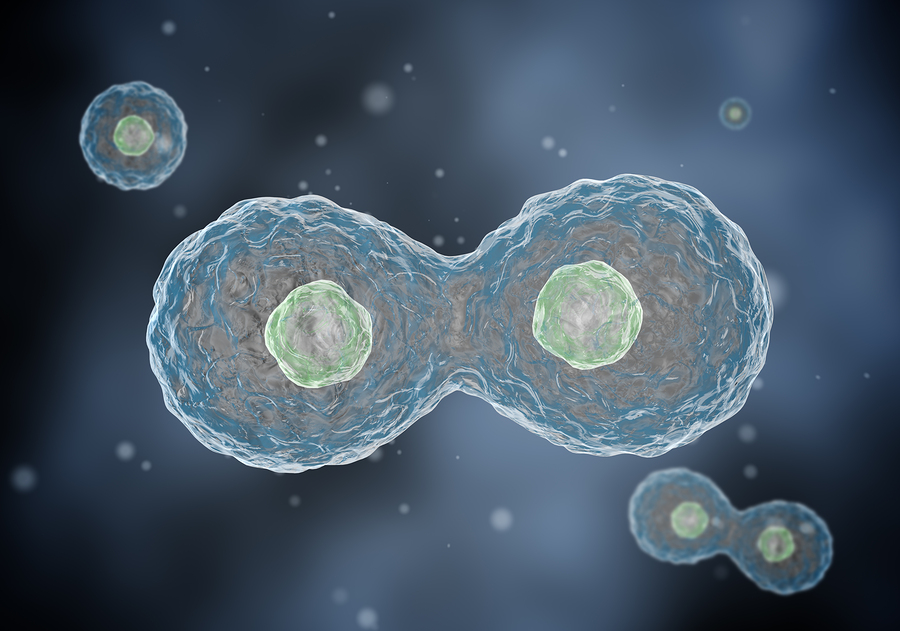
Is the size of a cell based on growth and division, its specific function, or is it regulated. New research shows that it is influenced by regulation as well as factors of growth and division. Most research is based on the idea that the longer it grows the bigger it will be, but this is not always the case. There are many other factors that affect size and growth. Cells can determine how they increase in size, but it is difficult to study metabolism and the feedback loops involved.
The size of a cell is determined by many factors—synthesis and breakdown, nutrition, environmental factors, protein production and autophagy. In some rapidly diving cells, including bacteria, the complex dynamic of growth and cell division determine some aspects of size. But, also, unique functions and physiology affect it. The type of cell that it is becoming from a stem cell—differentiation to a specific sized cell—also is influenced by multiple factors. Cell division cycles controlled by genetic networks, also, determines size.
There are many intersecting questions:
- Best size for the cell type
- Response from environment
- How do cells measure themselves
- How does the cell know it is a different size
- In the cell division process, how do cells change their growth to promote specific size
- How do size changes affect functions
- What is the best size for function
- How do cell sizes cause illness
- How do cell sizes cause aging
Organs Maintain a Particular Sized Cell

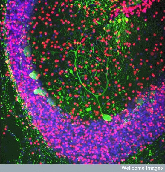
Every organ in the body, but particularly the brain, functions accurately because cells maintain a very specific size and shape. A lot is known about how cellular networks increase or decrease their growth, such as using the remarkable intelligent mTOR pathways described in another post.
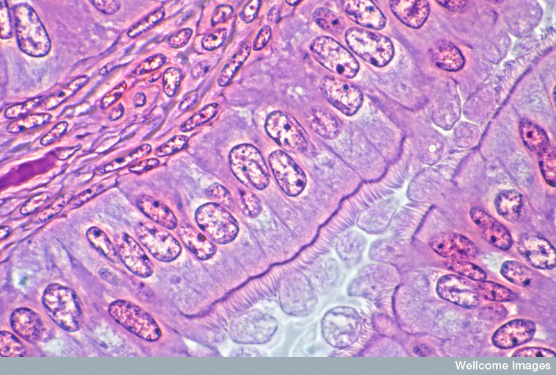
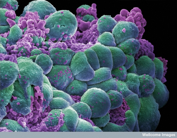
But, this doesn’t begin to answer the question of how the cell knows how it should fit into a larger vision of many cells working together at specific sizes. It is quite remarkable that in organs, most of the cells are the same size, but different between tissues. Epithelial cells are particularly uniform in shape and size and changes are often a sign of the development of cancer.
Even if we consider signals from other places to all the cells, there should be more variability. This variability is dependent on the way the cell’s divide, the rate of growth or nutrient availability. In order to maintain similarity of size with exactly the same environmental factors, there must be an active regulation. Individual cells must measure their own size and then regulate exactly how small or large they must be.
Cells Measuring Themselves
To determine how large a cell should be it, it must at least alter growth rates and the cell cycles. Different tissues have various abilities to maintain these appropriate sizes—some more effective than others. In many tissues, if cells are the wrong size they don’t perform well.
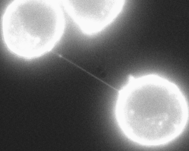
It is the number of cells, not the size of an individual’s cells that makes one person larger than others. Despite this, organs maintain exact standards of cell size even when people are rapidly growing. During the production of new cells from stem cells, they can become much larger, by orders of magnitude compared with the original stem cell. Bone cells increased their size by more than 10 times when the bone is growing. In the pancreas there are two different cells, right next to each other, where one is two times the size of the other. These examples show that size is highly regulated.
 Receptors on the cell’s membrane respond to many signals from the environment that adjust the size of cells—cytokines, growth factors and molecules that signal cell division. Some factors increase cell growth and other stimulate the cell division cycle producing more cells. Some of the same factors function differently for specific cells. Research shows that, although these signals determine the average size of cells, they don’t actually determine the variance of individual cells. This needs the ability of the individual cell to participate in the process. Is there an optimal size for cells to function?
Limiting Variations in Size
When cells are first produced from division, there is a time that they are growing when ultimate size can be determined. One way is to only have cells that have achieved the “correct” size to be able to divide and make more cells. Large cells divide rapidly to cut down the size of the cell in half. Research shows size measurement in the G phase of cell division.
The very complex cell cycle is the process where cells divide and make a new one. It is described as the interphase and then mitosis. The interphase is broken up into parts. The G phase is the “gap” between getting ready and then actually dividing. In G1 cells increase in size and are prepared for the very complex process of copying of the DNA.
The next phase is called S for “synthesis” of DNA. This is when the complex process where copies of the chromosomes are made and divided.
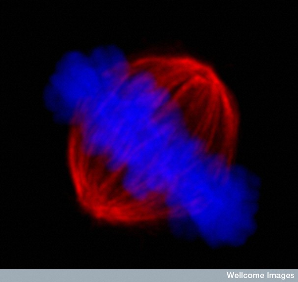
 G2 is the time between DNA copying and mitosis when the two cells are pulled apart by a complex microtubule driven process. G2 serves as a checkpoint for size and preparedness. In G2 the cell continues to grow larger. It is here that research shows some regulation of cell size.
In mitosis all cell growth stops and all the energy is put into the orchestration of the breakup into two complete cells. There is another checkpoint in the middle of mitosis (metaphase checkpoint) where everything is checked before it completes the division.
During the cell division cycles, research shows that cells in the S phase have a very variable age, but are very similar in size. In the G phase, cells stay as long as it is needed to become a particular size. There is a process during G phase that large cells become smaller.
 Its very difficult to measure how fast an individual cells grows, although there is recent progress with lymphocytes. This study show that the measurement that gates the G phase is consistent with control of rates of cell growth. Slow cells need to grow faster and when they arrive at a specific size and rate they are released. But, other studies show the opposite—the measurement is how large a cell has become.
It is, also, not really known whether cells grow linearly or by orders of magnitude. For the latter, larger cells would be growing faster. This would need another source of regulation. Although mathematical theory related to measurements of cell growth existed for many years, actual accuracy levels have only been achieved recently and, in fact, growth rates are more complex than either. The larger the cell the faster the growth rate, except for very large cells and all are faster than thought in the G phase. Therefore, a strong regulation has to be occurring from somewhere else. This research showed that the larger the cell and the older, the more they divide and make new cells.
Cells Measure Themselves Against an Ideal Size
Skin cells are produced in a very narrow range—between 12 and 14 micrometers. There are gradients inside the cell with less of a particular molecule that simulates cell division in the center of the cell and more at the edges. This gradient seems to trigger cell division at a particular cell size. It prevents the mechanisms of the dividing cell near the edges, keeping the cell above a certain size.
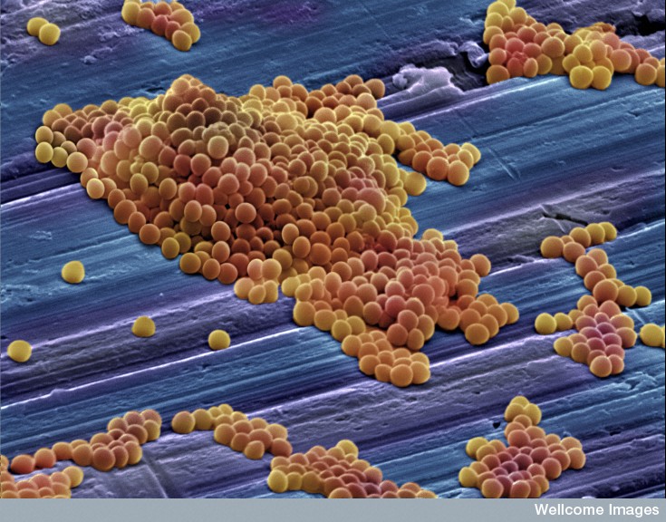
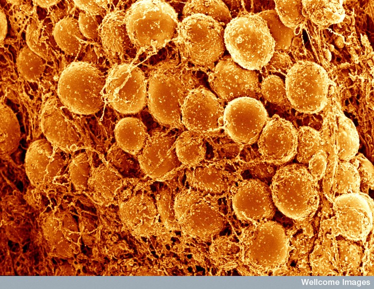
This mechanism has only been, recently, proven for a particular bacteria species. In this situation, the bacteria with a larger initial size divides earlier, whereas one with a smaller initial size divides later. There are transient oscillations, not just in cell size, but also in constitutive gene expression. The cell’s initial size determines how much it will grow before it splits into two.
Another possibility is from another bacteria that measures its own growth rate. A certain rate of protein production is necessary to get out of the G phase to the S phase of cell division. In the beginning of the G phase of activity molecular feedback starts size control. But, later a different mechanism occurs. Small cells take longer in the G phase. Size control is related to a molecule Cln3 in G. But later in G the size determines whether it can leave that phase. Small cells stay longer till they are a specific size and then they proceed to the next phase.
Cln3 senses the need for more protein production and, also, allows the exit from the G phase. However, when there is a very small production of proteins, no Cln3 is made. It is destroyed early and this has a direct relation to the rate of proteins appearing.
 Another specific molecule, also, determines the procession to mitosis in another situation. Cln3 operates through affecting the inhibition of transcription factors related to the cell cycle and producing a feedback loop involving Cln2 and Cln1. These start the complex cell division cycle
This mechanism uses a titration of a special molecule against a fixed number of binding sites on the transcription sites. The size of the cell determines this molecule. These molecules somehow trigger the G phase.
Yet another molecule, cyclin E, also senses the production of proteins. It, also, has a differential between transcription and translation which is used as a signal for translation.
Effects of Cell Size
 Different physiological situations determine whether there are more cells or larger cells, when more function is required. But, cells have many different functions and so this is hard to determine and there has been little research into this until recently.
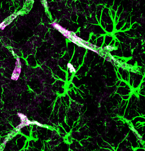
Neurons have to be a very precise size to fit in the circuits. Size of neurons greatly influences structure and function in the brain. But, in other organs is it possible that fewer larger cells can do the same job? This would mean the determination of the organ’s uniform cell size is not from the efficiency of its specific functions.
Many different situations alter cell size. In the kidney, the rate of liquid flow affects the size of the cells by triggering cilia sticking out of the cell into the fluid. Lymphocytes change size in response to cytokines. Microglia change shapes and sizes in response to inflammation. (see post)
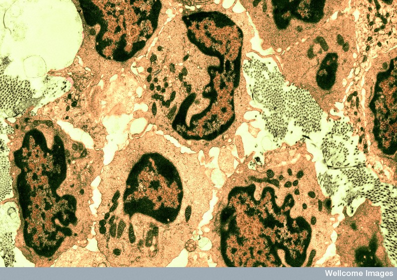
 In pregnancy, the pancreas increases βcell’s size to make more insulin. But, in diabetes, where βcells die, the number of cells increase, not the size of new cells. Liver cells, also, increase size in pregnancy, but after surgery the number increases instead.
The rate of insulin production in βcells is shown to be more connected with cell size than metabolic measures. Metabolism, insulin and protein production are all correlated with βcell size. In another situation, smaller cells produce less insulin. Exact mechanisms are not clear.
Fat cells have been studied, where large and small cells have different genetic networks operating. When a fat cell is larger, the relation to extra cellular matrix changes through signaling pathways that change transcription factors. The matrix might produce more regulation for the larger cells. The large cells have very different metabolism and make different products and have more synthesis of lipids. These types of changes occur, also, in liver cells and red blood cells.
 Large fat cells have different responses to insulin, sugar and fatty acids. These differences show that there is an optimum size for function. Larger fat cells produce more diabetes.
Cancer cells, also, have different behavior with different sizes. Larger cells are more malignant and have more differences between cells including varied shapes. These differences are not based on the environment, but are determined by the individual cell.
How Do Cells Know What Size They Should Be?
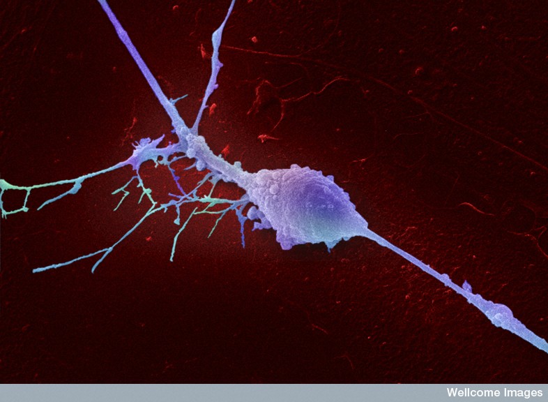 Cells understand quite a bit about themselves and their environment. It is remarkable what individual cells can do by themselves. They can send and receive wireless signals with many different types of cells and act upon the information received. An individual cell can understand where it is in relation to the entire body of trillions of cells. An individual leukocyte can travel to an exact distant location through many unusual and often unpredictable situations and terrains, while changing shape and methods of travel. An individual neuron in the fetus can travel an enormous distance to an exact spot amidst billions of cells. In the adult, axons do the same. Cells know what types of cells they are supposed to become. And cells know exactly what size they should be.
Where is the direction for the actions of this individual cell among trillions? How can anyone not be impressed by the intelligence of individual cells? Does this vast cellular intelligence show that the cell is connected with a form of mind?