 Many issues confront neuroscientists in their attempts to map the brain. A previous post, the Limits of Neuroscience, listed critical problems. Connections are made and pruned each day and one neuron can have 100,000 connections. There are many types of synapses, many types of neuroplasticity in wide networks, and many different types of postsynaptic densities (the complex structure in the post synaptic neuron with over 1000 large intersecting proteins.).
A major complication is that neuronal circuits fire in a millisecond, while the same neuron can participate in a totally different circuit the next millisecond. Neurotransmitter types are not static, and subunits of receptors can change, as well. Electrical properties vary, as do myelin patterns. Most mapping projects do not count the astrocyte network, which is not only bigger than the neuronal circuits, but controls the production and maintenance of synapses. Also, neurons participate in multiple types of communication—synchronous brain waves, nanotubes, exosomes, and electrical synapses—not just axon dendrite connections.
But, one of the biggest mysteries is the number neuron species and how to define them. How many different kinds of neurons are there?
How many Neuron Types
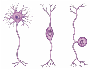
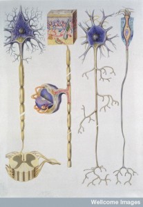 There has been no clear way to define the different species of neurons. Obvious categories include structural differences in shape and positioning of dendrites and axons. But, there are many more functional characteristics—different architecture, density, size, electrical properties, microtubule scaffolding structures, synaptic structures and circuits. Dendrites make different spatial and timing arrangements.
Previously, 40 regions were defined in the brain, each with different shaped neurons, perhaps a billion of each type. A very recent study showed that the genetic network differences are not in sync with these anatomical differences, which means that there are many more types.
It is not really clear how many neuron types there are, nor is it obvious what all the characteristics of different neuronal species are. Recent findings are demonstrating a much greater number of varieties.
 This post will look at recent research of the eye’s retina. Because of its very detailed structure, the retina has been, perhaps, the best place to begin to study this question.
The research findings don’t imply they will translate to other regions like the cortex. In fact, the vast and much more complicated cortex, may have orders of magnitude more neuron types. Very surprisingly, different cortex layers have been shown recently to have many different types of myelin patterns. So, different regions many be extremely varied in the types of neurons, as well.
Mapping the Brain — Neurons Develop At a Fantastic Rate
 In order to populate a trillion neurons for the full-grown fetus, near the end of fetal life, different types of neurons are being added at 250,000 per second. Later, the trillion neurons are pruned to just 80 billion—which happens fairly rapidly with no scarring—each neuron is taken apart by planned and well-organized programed cell death. These remaining neurons each produce up to 100,000 connections to other neurons creating the truly staggering number of 1015 synapses. It is not known how many specific kinds of neurons are created originally, or whether some develop variations after the pruning.
One of many brain projects attempts to map the mouse brain, which has a mere 70 million brain cells with 14 million in the cortex. The maximum number of connections for one mouse neuron seems to be 45,000. Mapping this much smaller number of neurons is currently an impossible, monumental project. If all of the existing supercomputers were used for a small region of the human brain, it would take thousands of years to calculate all of the possible connections.
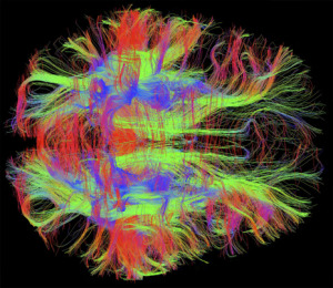 Current technology does not allow us to observe individual neurons while they are alive and active in the human brain. Millisecond action of brain circuits are not observable because MRI’s only generate average information of 70,000 neurons over a second time frame. Instead, to answer this question, very detailed work must be done to gradually piece together individual neurons activity, structure and function.
Study of the Eye
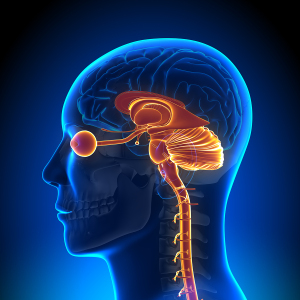 The retina neurons have been widely studied because the retina is highly structured in well-defined layers.
This post will describe the work in this one small region of the eye and the different neuron types in different layers. Even this small region has more than 70 different types and possibly many more.
But, in fact, it is difficult to even figure out what defines a category of neurons. Most studies are in the mouse brain, which is vastly simpler than the human brain. But, it is relevant as a brain of another mammal that has many common features. The analysis of what makes a type of neuron involves studies of neurochemistry, the wiring of circuits, the anatomy at small and larger scale, and physiological studies measuring metabolism of neurons and electrical behavior.
Mouse Retina Is a Good Place To Study Neuron Types
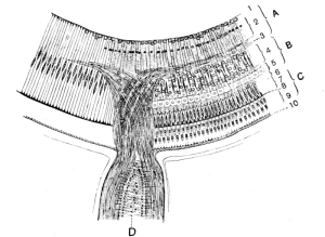
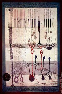 The retina is built in five layers—cell bodies in layer one (outer layer), layer three (inner layer) and layer five (ganglion layer). The two layers in between are composed of axons, dendrites and synapses—the outer plexiform layer (OPL) and the inner plexiform layer (IPL). The two plexiform layers of synapses are divided, also, into many sub layers called strata.
There are five classes of neurons, each with many different types: photoreceptor cells, horizontal cells, bipolar cells, amacrine cells and ganglion cells. Most of the diversity is in the bipolar, amacrine and ganglion types. All of these cells form connections with each other in the two layers filled with dendrites and axons.
In biology, it is not always easy to define species or types. For example, microbes have a vast amount of “species” of which we have barely scratched the surface. Types of neurons are defined as those with similar shape, structure, molecular structures such as the types of proteins used, electrical functions including the rates and types of spikes, genetic networks, the types of metabolic functions.
 Other characteristics include:
- There are neurons that have an on and off switch.
- There are neurons that express specific secretions and membrane bound receptors.
- Neurons have different orientations, shapes, and symmetries. For example, specific neuron types have dendrites that have a particular form.
 Also, in the retina different cell types connect in the many sub layers of the IPL and OPL. There are some with small amounts of connections in a sub layer and others with a large amount.
- Some cells have input and output connections, some one or the other.
- Some cells lack axons and have only small dendrite connections.
- Some have different spatial arrangements of dendrites.
- Different electrical properties
- Different neurons respond to transmission in different ways
- In the retina there are cells stimulated by light, others stimulated by darkness.
- There are cells that detect specific kinds of movement
- Diseases affect only certain cells
Unfortunately, these criterion don’t always work and there could be a lot more factors. Because of the difficulty in defining neuronal types, studies have not been prominent until now. When cell types are known, then the types of calculations and circuits can be discovered.
Bipolar Neurons of the Retina

The bipolar cells are made of a central body with two opposing processes. They take signals directly and indirectly (through amacrine cells) from the rods and cones. The photo receptors are stimulated by different types of light and send signals to the ganglion cells on their way into the brain. They, also, receive signals from horizontal cells.
Bipolar cells are divided into cells that respond to light and those that respond to darkness. There are at least twelve different types of bipolar cells. There is only one kind of neuron, it seems, that is connected with the rods of the eye, and up to 12 that connect with cones. These varieties are defined by anatomy, genetics, and the connections in the sub layers. By functional characteristics, several of these types may actually be multiple subtypes.
Each characteristic of the bipolar cell could conceivably be broken into more divisions. Two of the types have different tiling patterns and are therefore considered different. There are other overlapping functions. Another confounding issue is the density of the cells in regions. Some of the types appear to have specific densities, while others don’t. Instead of 12, there could be many more types.
Ganglion Cells of the Retina
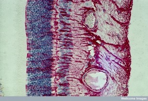 The retinal ganglion cell (RGC) is a neuron in the inner surface of the retina—a layer called the ganglion cell layer. Visual information travels to the RGC from amacrine cells and horizontal cells. They make complex connections with long axons to many parts of the brain—thalamus, hypothalamus and midbrain—with visual information that forms images.
These long axon bundles are called the optic nerve and the optic tract. There are many different sizes of these ganglion cells, and there is great variation in the amount of connections and what type of visual information they respond to. A previous post described a group of these cells that respond to light, but not for vision; these cells are part of the circadian system and the pupil’s light reflex.
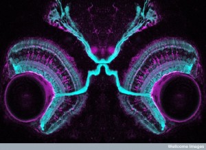 Ganglion cells go via the optic nerve to the brain. Each cell is thought to have a particular computation of a visual pattern. Knowing the types of the ganglion cell would describe what the eye is telling the brain it sees.
The very laborious task of mapping dendrite connections shows possibly 20 or more types of neurons, but it is difficult to be certain since this is such fine, laborious work. From a purely physiological view there are possibly four classes based on the direction that the visual information flows. Some cells only pick up front motion, some only side motion. When looking at the genetic networks of cells, more different subtypes were found.
Dendrites overlap and this has become a new criterion for neuron species. There is, also, the question of the periodic placement of the cells in space and their independent function. This technique also found many of the same subtypes. Genetically, many types are found on the basis of making particular molecules.
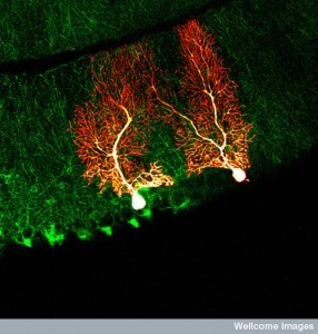 The density of the arbor of dendrites is considered, but this actually obscures the many specific differences—trees that can’t be seen from the forest view. Other factors have been the angles and tortuosity of the dendrite arbor.
It has been thought that the size of the dendrite arbor measures the amount of connectivity and synapses, but this is not necessarily the case. In fact, understanding the structure of the arbor doesn’t predict the relations between different arbors. But, when looked at statistically, this approach has been consistent with many of the cell types. Studies with electron micrographs have come up with 12, 15 and more overlapping types.
Amacrine Cells
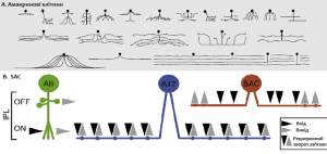 Amacine cells are interneurons that send 70% of the connections to the ganglion cells. The rest of the input to the ganglion cells comes from bipolar cells. They are found in the inner plexiform layer of the retina—the second layer with synapses of bipolar and ganglion cells.
Many amacrine cells don’t have axons, but rather sit nearby and influence the flow of information from bipolar cells (like horizontal cells). There are at least 22 different types of amacrine neurons, each performing many specialized tasks that connect with specific types of bipolar cells and specific types of neurotransmitters. One type connects the information of both rod bipolar cells and cone bipolar cells.
Amacrine cells have been more difficult to define and study than the other cells. At least 23 different types have been proposed by sparse labeling technques. But, in the electron micrograph studies there are at least 40 different amacrine neuronal types noted. The arbors of these cells are vast and studies of their connectivity is not yet able to be done. Also, amacrine cells are more sparse in the general anatomy of the eye making it even more difficult.
Neuron Types in the Retina and Elsewhere
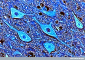 The retina is a sheet like structure with definite layers and therefore, the neuron types should be easier to define than other more amorphous cellular structures such as the vast cortex and cerebellum. When we refer to the location of the neuron, usually it means the cell body. But, the more relevant consideration might be the details of the axon arbor and the dendrite arbor.
It is not clear that there are easily definable rules for mapping the connections of the brain. Cell types are usually defined by the anatomy of single cells. But, they could also be defined as connectivity patterns.
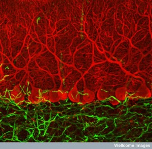 Can the retina help understand other areas of the brain such as the cortex? The retina is highly organized, with the cortex more diffuse. The cortex has layers, but not definite synapse strata as the retina. The recent studies on myelin show that different cortex layers have different patterns, which recently more complexity. (see post). Each of these myelin variations are probably different neuronal species.
The splitters define many species and the lumpers few. Is it possible that there is a continuum of neuron types?
In the cortex, first there were pyramidal and non-pyramidal neurons. Now, there are at least 50. It is possible that there are more than neuron 500 types in the cortex.
How Many Kinds of Neurons Are There
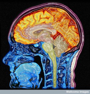 With a fantastic amount of different factors, study of neuronal species has to begin somewhere. Studying one small region of the retina shows over seventy types of neurons and perhaps many more–40 amacrine, 20 ganglion and 12 bipolar. This large amount of varieties is found even before we can visualize and study individual neurons.
Originally, there was believed to be 40 different brain regions, each with different neurons. In the cortex, for example, early research decided there were two types, pyramidal and non-pyramidal. Then, more than 50 types were identified. Now, possibly more than five hundred cortical neuron types in that one region. The retina, a much smaller region, has more than 70. If many small regions have 70 types, there are most likely thousands in the whole brain, each with different properties.
Is it possible that each neuron is an individual and slightly different? Is there a continuum of types? Can new types evolve and grow in individual brains? These are the many unknown questions that will need to be answered to map the brain.
There has been no discovery of a central place for mind in the brain. The increasing number of neuron types greatly complicates mapping the brain to explain mind. Perhaps, this is because mind is interacting with and giving direction to each of these unique different neuronal types.
