 The brain is extremely dynamic, building and pruning connections in milliseconds with many different types of neuroplasticity simultaneously arising in large circuits all over the brain. The holy grail of neuroplasticity has been the creation of new brain cells in adults. Research looking for one cell in a region of the brain is much more difficult than a needle in a haystack. Despite, overwhelming odds, study shows new neurons arising in at least three, and possibly more, places in the adult brain—both humans and other animals.
Recent research shows that new neurons at least in the critical memory center of the hippocampus, are essential to learning and memory and rapidly become part of existing circuits. In fact, they remodel the circuit to take in new material in new ways. A previous post described that more than a thousand different types of neurons exist. Neurogenesis research shows that these new adult born neurons may be completely new types of cells with increased excitability, more widespread connections, a new window of opportunity, and increased neuroplasticity. New neurons in adult brains remodel memory circuits to make information more specific.
 A previous post mentioned many triggers for the new cells. Exercise, psychotherapy, treatments for depression and enriched environments for learning all increase the rate of production of new cells. In depression all three types of therapies increase brain cells—medications, psychotherapy, and lifestyle changes. BDNF (brain derived neurotrophic factor) and steroid hormones are important triggers for the new cells. One confounding factor is that only some of the research has been able to be done in humans and human neuroplasticity and neurogenesis might be quite different than other animals. There is wide variation of new cells in different animals, for example between rabbits and mice. One fact in humans that is encouraging is that the entire hippocampus dentate nucleus is rewired with new cells in the adult, making this critical memory center focused for adult life. This is one way that the elderly brain is better than the young (see post Does Cognitive Ability Improve in Old Age?)
New Neurons
In the fetus there is massive creation of new cells. At one point in the third trimester, 250,000 new cells are born each minute, making the total near a trillion cells. Later, these are pruned to 80 billion and trillions of connections are formed that are, also, later pruned. In adults, new cells are definitely found in the hippocampus and in the SVZ (subventricular zone). From the SVZ they migrate along a rostral migratory stream to other regions. In mice, they are then found in the olfactory bulb related to smell cognition and memory. In humans, they are found in the striatum, a major center for learning, memory and movement. Another study found them in the limbic system in adolescent hamsters. These cells were integrated into circuits related to social learning, evaluation of facial expression, body language and mating behavior. New cells were, also, found in the hypothalamus, stimulated by fat cells to coordinate metabolism.
In the hippocampus, the new cells have been shown to respond to the new incoming data, and may actually replace old memories with new ones that are more specific. They are clearly correlated with new abilities to separate and discriminate memories, called “pattern separation”. Another form of memory, which detects similarities, called “pattern completion” appears to be maintained by the older cells, until this is replaced with more specific details.
Another finding has been that exercise opens a window of neuroplasticity where more new cells are created. This can be for good or harm since one study showed that during this window addiction occurs faster. Also, in sleep there are more cells. Only in humans is there a slight decrease of new cells with aging. Cells occur near blood vessels and one blood borne chemical that increases new cells are CCL11(CC chemokine ligand 11). A study recently showed that a decrease in the factor was critical for the decrease in new cells in the hippocampus with aging.
Many different neurological and psychiatric diseases are now known to affect the production of the new brain cells—reductions noted in mood disorders, addiction, stress, depression, epilepsy and Alzheimer’s and Parkinson’s disorders. Cures of some of these—mood disorders—are associated in increased production.
What Can the New Cells Do?
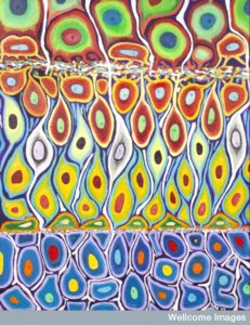 What effects do the new adult brain cells have on brain circuits? Stem cells make new brain cells in specific regions of the brain, which transform into neurons and travel to integrate into brain networks. The best studied regions for these new cells are the subventricular zone (SVZ) near the lateral ventricles and the subgranual zone of the dentate gyrus (DG) of the hippocampus.
The SVZ cells become part of the first central relay of the olfactory bulb (OB) as GABA interneurons. These new cells appear to be critical to maintain the basic structure of the olfactory bulb in mice (not clearly in humans). The hippocampal cells are glutamate granule cells.
The new cells appear to have different properties. They are more excitable and have different kinds of plasticity and connectivity. More recent research shows that entirely new types of neurons are made. In fact, it is not clear how to define neuron types since there are so many variables. Please see the post, How Many Kinds of Neurons Are There, for discussion of the thousand plus varieties.
New cells in the brain soon receive already mature developed inputs and top down well-developed systems.
Circuits Want More Cells
The adult hippocampal neurons deal with time and space memory and olfactory neurons with smells. Both analyze and respond to detailed information during their entire lives, often many decades. There are a host of glial cells that have supported these neurons in their complex tasks, by pruning dendrites and supplying neuotrophic substances that nourish the neurons.
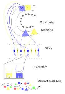
New Smell Cells: In the olfactory bulb, almost all of the newly born neurons become active functioning GABA cells in about a month. Even as the baby cells travel in the migration route, called the rostral migratory stream, they have already made many functioning receptors for NMDA glutamate and GABAA. These cells can already respond to the signals that occur in the regions they are travelling through, mainly the olfactory cortex.
In the bulb, synapses form rapidly for the GABA and glutamate neurons. They are positioned in the region of the bulb that has the most top down signals from all over the brain, including cortex, limbic and subcortical. This information from far regions of the brain sculpts the early neuron’s function. After several weeks in position they start forming larger dendrites to the external plexiform layer to receive connections to the mitral tufted cells (M/T). They first have connection the upper cortex then only the local sensory information.
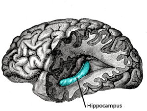 Hippocampal New Memory Cells: When the young hippocampus cell first arrives, it receives mostly local inputs from GABA interneurons, which are inhibitory. They have more excitability that the older cells and are more responsive in, at first, sensing only the local environment. This inhibition was involved in regulating the stem cells that created the new neurons.
Several days later other local excitatory mossy cells connect. Only later do the cells connect with the inputs from the entorhinal cortex, when more complex feedback loops occur with the inhibitory interneurons. As the neuron establishes more connections, astrocytes gradually connect to the synapses. Microglia start to eat cells that don’t make it, which stimulates more cell production.
Activity influencing New Cells
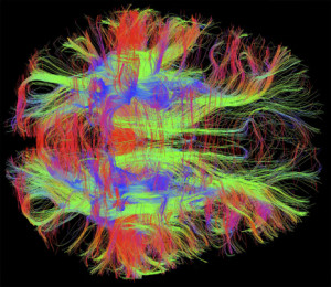 A previous post noted that activity doesn’t always produce increased connectivity or pruning. Individual neurons can act quite independently. But, in the smell circuits, research with increased and decreased levels of smell sensory information from the environment does lead to more cells and more survival of cells and more synapses. Another factor is the excitability of the cells. Those that have greater excitability can survive in in situations of deprivation of sensory signals
New cells respond to signals from the local or top down neurons in a critical time window of up to a month for survival and several months for making new synapses. The activity increases when learning or reward effects are involved. When activity of the animal being tested is increased with exercise, enriched environments or new learning, then more new cells are taken into the circuits and survive. This enriched activity increases connections both locally and top down circuits.
Timing of Synapses with New Neurons
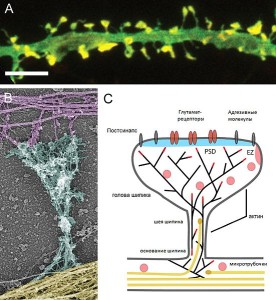
New neurons take in information before they begin to send out signals, although even when they are first travelling to their position they send out GABA neurotransmitters without any synapse. This might be stimulating to the circuit as a whole. This type of behavior is unique to the adult born neurons rather than those born in the fetus, which possibly makes it a unique type of neuron.
In the hippocampus, synapses are prominent three weeks after birth of the new cells. These cells develop synapses to inhibitory interneurons and provide a hyper excitable inhibition to the entire circuit at two months, while they themselves are not inhibited. These new cells, therefore, influence the entire circuit and region. They have a wider range of response that the older cells. They are more active with new information, such as new smells, and they are particularly sensitive to reward based memory. These new neurons, therefore, mold the entire sensory memory system. These cells, also, provide more temporal integration and pattern separation.
Inhibitory interneurons normally provide many control functions. But, these new cells are glutamate type and not interneurons but they shape the entire circuits, as interneurons normally do. They appear to attract inhibitory type neurons.
New Neurons and Pattern Separation
 New neurons have a special ability to increase a type of learning called pattern separation. Pattern separation is a process that makes similar memories and patterns more distinct and specific. It separates parts of memories into very specific details such that they do not overlap. It involves storing similar information with very specific differences.
These new cells appear to provide pattern separation for information that was previously confusing both the hippocampus and the smell centers. These new neurons born in the adult brain are needed to make distinctions for spatial and visual information. They are especially important if the task is more difficult and ambiguous. The old and new cells have different roles and ratios are altered, which is important. While the new cells do pattern separation, the old cells maintain less specific older memories called pattern completion. Pattern completion occurs when the new neuron is hyper excitable, forming connections, and altering the entire circuit. It affects the plasticity of the circuits forming new types of synapses that didn’t exist before.
In the smell centers, the hyper excitable cells increase inhibition of the out going signals, which makes it possible to distinguish the details more.
In the hippocampus it is more complex in that the cells attract the inhibition that helps the pattern separation. The new cells take over the transmission of the data from the region to other centers thus influencing the specific nature of the signal. The GABA inhibition lowers the activity of the older neurons, changes the inhibition and the network.
Both Types of new cells Help Learning and Memory
Hippocampus: In the hippocampus, spatial learning and associations of space with other aspects of experience such as emotion are increased. If the new neurons are damaged early this doesn’t occur. But, if damaged after months the effect has been generalized and it doesn’t affect it.
The new cells separate out specific circuits to define specific patterns along the lines of pattern separation. The new cells affect the “sparse coding,” that is, they only allow very specific memories to form. And when the detail is remembered it needs to use the new neuron. This process allows for the specific memory not to damage or interfere with existing memories. The more the memory is separated by time, the more new neurons are needed. The new cells are necessary for the new memory during the cell’s stage of increased excitability. These memories are then sent to the cortex for consolidation of the memory. (Every time we re-remember something, there is a period of time for consolidation that can be altered – useful to alter the effects of traumatic memories).
These new cells are particularly connected to the hippocampus region of social memory (CA2). They are involved in both cognitive (dorsal) and emotional (ventral) information. Separate circuits from distinct regions of entorhinal cortex connect to the hippocampus. The purely spatial cognitive tracts have many new neurons dealing with exploration of space. The emotional circuits from the amygdala, pre frontal cortex and hypothalamus have less new neurons that more slowly mature related to anxiety, fear and stress. These new neurons, also, are less excitable. They are, also connected to the cortisol hormones.
Most studies of new cells have been about memory. Having a large number of new cells can affect previously memorized incidents. New neurons can increase forgetting of past memories. One study where mice learned a new skill, new cells were very increased. When the new cell production was artificially stimulated they forgot much of the material. Therefore, having a great amount of new neurons can eliminate previous old memories. It is theorized that it increases transfer of the memory to a more permanent status in the cortex. Therefore, the new cells may clean up memories waiting to be consolidated.
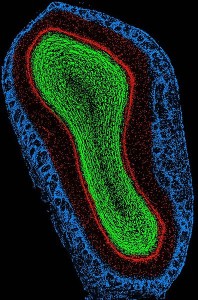 Olfactory: In the olfactory region new learning increases by the new cells making stronger synapses with older cells, creating a plasticity from the new to the old cells.
The olfactory bulb has unique ability re model circuits using new cells related to reward. This circuit alteration occurs in the cortico-bulbar tracts. This specially occurs during deep sleep where top down signals are specially connected with these new brain cells. Response to food and hunger are, also, connected in this way to olfactory smell cells. Therefore, new cells are connected to both new sensory information but, also, complex internal states such as stress, sleep, reward, motivation and attention. These complex factors sculpt the effect on synapses and circuits of the new cells. They connect the message and its context.
Different animals, such as mice versus rats, have different results in the olfactory. And it is not yet clear what occurs in humans. There have been conflicting results in the effects of stopping new adult born neurons in mice in very specific cognitive functions, such as following cues and spatial learning of mazes. Pattern separation was impaired in discriminating about context, but not a particular maze test. This shows that these new neurons are influenced by different factors including stress and hyper alertness.
In Humans
New cells appear to have many unique qualities in humans. They have increased connectivity, neuroplasticity, and excitability. Research in humans reveals that just about every neuron in the dentate nucleus of the hippocampus is turned over in adulthood. This occurs even though there is small decrease in the production of new cells in extreme age as compared with mice.
In the olfactory system, the migration pathway of the new cells is quite different in mice and humans. The entire organization of the smell glomeruli is very different in humans.
New research shows that new neurons travel and integrate into the striatum, near the SVZ, critical to cognition and movement. These appear to be inhibitory interneurons, which are the type lacking in Parkinson’s patients.
In humans, pattern separation is involved in memory, learning and mood. New neurons are needed for specific control of mood states. Lack of new cells in the hippocampus is correlated with depression and smaller brain regions and successful treatment with antidepressants with increased new cells. Lack of cells doesn’t allow for the critical distinctions of threats and safety. Discrimination ability in mood is altered with increased anxiety and posttraumatic stress.
Inflammation affects new cells with many microglia in the regions of new neurons. These microglia prune defective cells allowing only the strong to survive. Microglia can help or hurt the process (as with other inflammation processes – see post on microglia). New neuron add units to the circuit from the cortex to the cranial nerves and the hippocampus. They have connections to both local interneurons and distant cortex structures.
Stress and New Cells
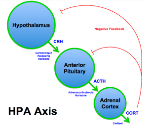 Adjustment to stress is very complex. A certain amount of stress is necessary for accomplishment and can increase new learning and new cells. But, toxic levels of stress do the opposite by decreasing new learning and new cells. These pathways are extremely complex and just being discovered. Steroids are the major stress hormones and are highly regulate by signaling between many types of immune cells and neurons. These involve morphogens from fetal development, neurotrophic factors, neurotransmitters and cytokines.
New research shows that chatter between cells that affect inflammation cytokines and glucocorticoid receptors have a big influence on new brain cells. In one study, when there was no cytokine IL-1R1 then there was no effect on steroid levels and no reduction in new cells in the hippocampus. This implies that IL-1beta is critical for the effect of stress. Chronic stress uses IL-1beta as a signal to the steroid receptors to reduce new cells. It is more complex because NF-kappaB and GSK3beta are also involved in chronic stress. Cross talk with these two are part of the IL-1Beta reaction. This signaling is very complex and, also, regulates the morphogens that create the fetal brain with new cells.
Steroids can regulate BDNF in different ways in different organs and cells. BDNF is highly involved in all of this signaling and is critical to form new dendrites. BDNF in turn, also, modulates glucocorticoids through complex feedback loops.
The effect of stress, and these signaling pathways, affect the circuits involved with the experience of good and bad experiences and in this way may be causative in depression. The complex interplay of the new cells and remodeling circuits is highly connected to these complex acute and chronic stress pathways.
New Neurons in Adult Brains Remodel Memory
 Brilliant research is able to identify the creation of a small number of individual cells, even in humans, and follow them to their integration into brain circuits. These cells, minted from stem cells, know where to go to integrate into circuits. The new cells appear to have unique qualities and are able to continue to transform, remodel and upgrade the human memory and cognition system.
These new unique neurons remodel our learning and memory. A previous post noted that elderly brains are superior to younger brains in all ways other than name recognition. The ways people use their brains creates new unique circuits and increased neuroplasticity. New more capable neurons transform the most fundamental cognitive centers. How we remodel them is our choice. Conscious choices and behavior affect the future structure of our brains.
Where is the direction for all of this? How does mind cause specific structures to be built?
