 When the topic is color a group of neurons oscillate with synchronous beta waves between two brain regions.
When the content changes from color to orientation a different group of neurons have the same synchronous beta waves between two other regions.
In this experiment it appears that synchronous waves are communicating information between two distinct brain regions with two different sets of neurons oscillating together at a specific frequency.
The two major theories of how the brain generates the mind are the neuronal connections where electrical signals travel along axons triggering a chemical connection at another neuron’s dendrite (see post Connectome) and electrical brain waves, which oscillate together at specific frequencies (see post Brain Oscillations). Both of these mechanisms occur simultaneously, so, perhaps they are complementary and perform different functions. Another theory of mind is that it consists of information, possibly in the form of electromagnetic energy, which would encompass all forms of electricity in the brain.
 Perhaps the neuronal synaptic connections are involved in computation by summing the inputs in the network of connections arriving at the dendrite. Perhaps the oscillating brain waves are binding together information from specific regions.
Both utilize electricity in different ways. In fact, there are many different sources of electricity in the brain.
If consciousness in nature, and the human brain, is in the form of information, electromagnetic or otherwise, it is not clear yet how this could work.
Where does brain electricity come from and what does it mean?
Measuring Electricity in the Brain
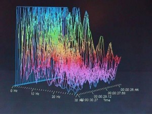 Current methods of measuring electricity in the brain utilize probes either on the scalp (the least intrusive method), below the skull coverings such as the Dura at the edge of the brain, and deep inside the brain. None of these measure individual neurons (which takes very advanced research techniques and is not generally practical), but rather account for the electricity in an entire brain region.
Recording from the scalp is called EEG, or electroencephalogram.
Recording below the Dura, one of the three covers of the brain, is called ECoG, the electrocorticogram.
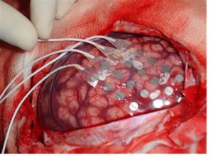 Measuring deep inside the brain with tiny electrodes deep is called intracranial EEG.
Measuring a magnetic field is called the magnetoencephalogram or MEG.
What is not generally known is that electrical measurements in a region do not measure a specific detail, event or component. All of these devices measure total voltage at specific points in a region, the addition of all electric potentials.
It is popularly thought that the major electricity in the brain consists of neurons’ electric signals along axons to the synapse to another neuron. This electrical signal, called the “action potential” travels along the axon and usually triggers the delivery of a neurotransmitter to another neuron. Occasionally this signal sends a more direct electrical signal through a “gap (electrical) junction,” to another neuron. The triggered neurotransmitter signals become part of a computation of all the signals landing on one dendrite from many different neurons. The summation of all of these signals on the dendrite of a postsynaptic neuron determines whether the next neuron will send a signal or not.
But, in fact, there are many other sources of electricity in the extra cellular space around the neuron.
First we discuss the well-known action potential, then the less understood electricity in the extracellular medium, which arises from many sources.
The Best Known Electrical Event – The Neuron’s Action Potential
All animal cells maintain a small electric charge with more negative charge on the inside of the cells. In most cells this doesn’t change. In the neuron there is a dramatic change when the neuron fires.
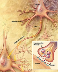 The action potential, sometimes called a spike, is a brief electrical signal, or current, that occurs in neurons, muscle, endocrine, and plant cells (a version can also occur in a bacteria to send a signal to the cilia to move). This is often referred to as the neuron “firing”.
Embedded in the membrane of the neuron there are a series of protein channels that allow small charged sodium and potassium ions to travel in and out in an orchestrated pattern down the length of the entire axon (which in the foot can be over a foot long). This occurs by the protein channel slightly changing shape. When a certain threshold is reached (usually 15 milli volts above the 70 mini volt resting state) then an all-or-nothing action potential is triggered that will travel the length of the axon.
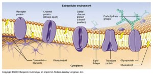 The charge travels along the axon “wire” by the coordinated changing of ion channels. By allowing more positively charged sodium ions through the membrane into the cell, these channels create an electrical charge, a voltage, when comparing the inside and outside of the membrane. The ion channels open one at a time along the length of the axon so the voltage moves from point to point all the way along the axon. Soon after the sodium comes into the cell, another channel opens to send potassium ions out to neutralize the charge. Then they gradually go back to the resting state.
The axon, the dendrites, and the cell bodies all have different electrical properties of their ion channels. Some areas can transmit action potentials, others cannot. The beginning of the axon, the axon hillock, is the most excitable. The cell body and axon are also quite excitable. Many neurons routinely spike at a specific frequency of 10 or 100 times per second, and then increase this amount with a given signal.
After the firing, which is referred to as hyperpolarization, there is an inactivated state, called the refractory period or “after hyperpolarization.” Another action potential cannot occur until this refractory period ends.
The same action potential mechanism also occurs with calcium channels in other regions of the neuron. But, the calcium channel signal lasts much longer, 100 milliseconds for calcium compared to 1 millisecond for the sodium action potential.
 There is one other very important factor in propagation of electrical signals – myelin. Myelin is a fatty substance that is secreted and maintained by a glia cell along many of the axons (Schwann cells in the peripheral nervous system and oligodendrocytes in the center nervous system). The sections of the axon that are myelinated do not have an electric potential; they are insulated by the myelin. There are breaks in the myelin at regular intervals, nodes of Ranvier, that are electrically excitable, like mini axon hillocks, and the electrical signals jump between these nodes travelling much faster than they would along an unmyelinated axon.
Local Field Potential – LFP
A different important measurement of electricity in the brain is the Local Field Potential or LFP.
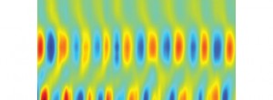 The LFP is the sum of all electrical gradients in a region of the extracellular medium, the space between the cells, filtered to avoid dominance by any one spike. An electrode that measures the voltage or electrical potential gradient measures it far enough away from any particular neuron to avoid one neuron dominating the result. It is thought that the LFP represents the synchronized input in the region, the sustained currents in the region from the soma and dendrite, rather than one specific spike output. The LFP is basically about the behavior of the entire group of neurons in the region.
It is not widely appreciated that if all the electrical events add together to form the electrical gradient at that moment, then the longer an event lasts the more opportunity it has to add to other signals. A very brief signal has to be very large to be measured in this process. Many small signals that last a long time will add together into a bigger voltage. This has been shown by the fact that a very small transcranial stimulation current can have a major effect after a while. A small brief signal without other signals at that millisecond will not add to anything else and therefore will be missed.
It was mentioned that the calcium action potential, or spike, is 100 times longer than the sodium. Because of this, the calcium spikes have 100 times more opportunity to add to other local events. Also, the long calcium spike can also be a trigger for multiple sodium action potentials where the sodium electrical signal will last a long time also.
Because of the complexity of the factors noted below determining the LFP, large events can be caused by the summation of many different events. An example is a theta rhythm in the hippocampus versus the neo-cortex, which might be assigned a specific cause, but in fact can be caused by multiple different very small events in the region.
 All electrical currents in a section of the brain combine to form the voltage potential that is the sum of all the activity. This activity is different at each point and forms what is called an “electrical field.” The electrodes measure this field in the range of milliseconds.
Any membrane, on dendrites, spines, soma (the cell center), axon and axon terminals, has an electrical gradient and contributes to the current in the extracellular medium. Glia cells often have slow moving gradients that contribute as well. In order for a small gradient to become measurable it has to be slow so that it will add with other gradients.
Synapse Activity Contributes to Extracellular Electric Fields
Synapses contribute much of the electric gradients in the LFP.
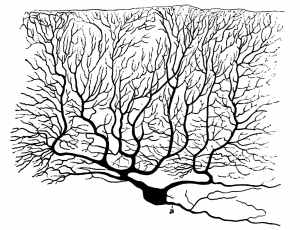 The dendrites are in the form of a large tree with thousands of branches with a membrane that is an insulator, unlike the axon where the electric current travels on the membrane. The large amounts of dendrite connections form a current inside the neuron membrane consisting of tens of thousands of synapses.
An example is the excitatory glutamate receptors where sodium and calcium ions flow inward. This creates a low level of these ions in the extracellular space
Inhibitory GABA neurons add a different type of current.
There are other channels in the membrane that are not related to the synapse action. Intrinsic currents, called Ih currents, occur by the action of other gated channels in the membrane (induced when the membrane is de-inactivated ) and It currents, induced by hyperpolarization of calcium currents triggering burst firing.
Resonance Factors in LFP
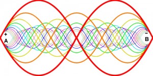 Several different types of neurons’ membranes respond to specific frequencies more than others. When this resonance frequency occurs it can cause much larger waves and it can trigger a continuation of the oscillations.
Theta resonance has been described in some cortex regions. In contrast some inhibitory neurons have been noted to have a resonance in the 30 to 90 ranges.
These resonance effects are influenced by the size of the triggers and the specific frequencies. To be large enough to influence the ECF potentials the same resonance has to occur in multiple nearby neurons.
After-Hyperpolarization Currents
 When an action potential occurs (the neuron “firing”) the electrical gradient of the membrane is called “hyperpolarized”. Hyperpolarization can be triggered by different ions such as calcium.
A factor related to calcium induced hyperpolarization involves the phenomenon of bursts of fast spikes and dendrite spikes. The electrical contribution of these bursts of spikes can be as great as synaptic events. These events can also occur in a coordinated fashion and can occur in greater amount when the brain is surprised by an unusual stimulus.
Electrical Gap Junctions – Electric Synapses
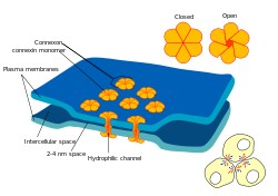 A gap junction is an electrical synapse, which is very small 3 nanometers (synapses that use neurotransmitters usually are 20 to 40 nanometers) and is very fast and often bidirectional. These synapses don’t amplify the signal the way other synapses may do with addition of multiple inputs. With the very rapid response these are often in rapid loops for rapidly needed defensive measures.
Electrical synapses can be involved in triggering synchronous waves and can cause increase in the currents of the extracellular medium, the LFP.
Glia
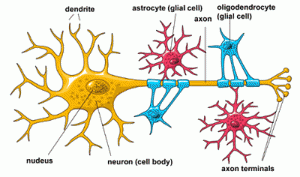 The complexities of the glia are just beginning to be appreciated. Other posts have described how glia are involved in monitoring the use and pruning of synapses. They are now known to provide many neurotransmitters to synapses as well. Activity in glia have major contribution to the electrical currents in the LFP.
Ephaptic Effects
Ephaptic coupling is a form of communication in the nervous system that is different from axon potentials and from electric gap junctions. It includes different ways that neurons may communicate including either very close physical contact of two neurons, or it may be an extracellular field effect that connects two neurons. It can have great influence on either action potentials or the LFP. It is one of the many unknown but important aspects of the electrical influences in the brain.
Neuronal Geometry is Critical LFP Factor
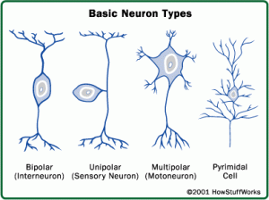 The most important factors in determining the extracellular field are the shape of the neurons and the rhythmic synchronized firing.
The shapes of the neurons effect the electrical contribution to the field. The most common cell in the cortex is the pyramidal cell with long, very thick dendrites that create strong dipoles causing a flow of ions in the extracellular space. Also, they are arranged in parallel columns with outputs at 90 degree angles, which causes these dipoles to be additive. Other animals have different amounts of this type of cell, different sized cells, and different angles that change the electrical properties. For example, the mouse LFP patterns have greater voltage than the rat.
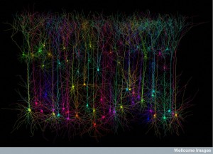 Circular symmetrical neurons that connect the thalamus and cortex have many dendrites of equal size in all direction. These create an isolated charge that is not additive.
The folding of the cortical gyrus create different electrical effects. On the concave side a gyrus pushes dendrites together. This curve effect on electrical function is dramatic in the hippocampus dentate nucleus.
The inhibitory neurons can create different electrical events such as gradients between a soma and dendrite in a particular neuron, and “inhibitory dipole.” For example, exciting a dendrite and inhibiting a soma cause the same electrical event and can therefore add together in the extracellular space.
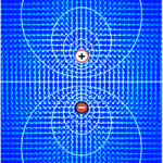 This can be confusing because a strong inhibition can create extracellular voltage but eliminate a strong firing of action potential decreasing the voltage. So, the LFP and spiking behavior can be very different even in a very small region. As mentioned in a previous post (the Connecome) the fMRI measures blood oxygen not electricity. Therefore, LFP in the gamma range can effect spiking and effect the interpretation of the fMRI.
Temporal Factors Critical in LFP
As well as architecture, an important factor in creating the LFP is the timing of firing.
If only geometric facts would be important then the cerebellum, with very ordered large Purkinje neurons, should have large LFP. But, most activity in cerebellum is local computation with small voltage. In circumstances where the cerebellum participates in synchronous activity then LFP can be much greater.
 The timing of the firing is very important in how long the voltage with add to other signals creating a larger voltage. For example, a slow frequency oscillation can influence a faster oscillation through a form of resonance called “phase-amplitude coupling.”
The specific local qualities of the extra cellular medium have effects on the conductance and transmission as well. Because of the complexity of the LFP a simple comparison of gradients across membranes of neurons does not give much information.
Electricity in The Brain and Mind
A previous post has discussed the research that supports the notion that synchronous brain waves appear to correlate with mental states. Neuronal action potentials clearly are involved in a process of signaling information throughout the brain that are related to mental states. Now, there is reason to believe that background, general inter cellular electric gradients can also be correlated with mental states.
Electrical currents include synapse effects, calcium spikes, action potentials, and the after potential of the spike. Most importantly, the specific neuronal architecture and the rhythmic timing affect these currents. Gamma rhythms can trigger other regions to oscillate and fire. Currents are affected by slower background rhythms throughout the brain. Because of the additive effects of longer events, small effects on the currents can have major effects. The general electrical background can affect individual spiking neurons through all the effects mentioned.
Although very difficult to obtain, a large amount of data about LFPs from the entire brain could produce a considerable amount of information about thought. In support of this, certain cognitive tasks correlate with specific types of LFP’s in various brain regions.
How these currents throughout the brain are related to the computation effects of the spikes between neurons, and the synchronized oscillations of groups of neurons between regions is not known.
 How does this all relate to the mind? It does appear that when different types of thought occurs, there are synchronized oscillations of groups of neurons between regions. Information for some type of mental computation is relayed throughout the brain by electrical signals and synapses. Also, specific cognitive events have specific patterns of electrical fields in the extracellular regions from the multiple sources discussed here.
It is tempting to consider mind in the universe as a form of information, possibly electromagnetic energy. There appear to be many different complex types of electrical events in the brain that correlate with mental activity.
This is still a long way from our understanding mind as an electromagnetic form of information.