 When animals perform an action in pursuit of a goal, specific brain regions appear to be activated. When animals observe that action, different brain regions are used. Twenty years ago a type of neuron was observed in the macaque monkey that appeared to respond in both of these situations. Later, these were shown in rodents and birds. Although there is controversy about whether these mirror neurons exist in humans, there is now one study that claims to prove that they do. Previous posts have shown that it is extremely difficult to study individual neurons in live humans, and can only be done prior to brain surgery with the cooperation of a patient whose skull is open. The limits of MRI have been discussed showing the dot of light, the voxel (the pixel of MRIs) measures activity of blood flow in a region of 80,000 neurons, far different than one neuron. (see post Limits of Current Neuroscience) Almost all of the studies of mirror neurons have been done with MRI, which only shows these large regions. Certainly, many of the popular theories, which place mirror neurons as the basis of human empathy and language, have yet to be proven.
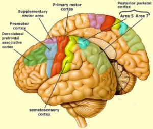 Even without proof of the existence of specific neurons in human beings that serve this function, there are clearly regions of the brain that do. These regions have been found in the premotor cortex, supplementary motor area, the primary somatosensory cortex, and the inferior parietal cortex. The one study that appeared to show human neurons with mirror properties involved 21 patients who were treated for severe seizure disorder. These patients were awaiting brain surgery and allowed intracranial depth electrodes to identify seizure initiating regions.
Doubts
 There are many that doubt the existence of specific neurons of a distinct class for this purpose. These scientists would assume that regions of neurons that provide a variety of functions might serve a similar function. Another complication is that the neuron would have to activate only if it could understand the goal or purpose of the action, which would require other brain regions in a circuit. It is, perhaps, too much to assume that a neuron could understand the intentions of others. If this were true then an individual neuron would, in essence, be thinking.
Macaque Mirror Neurons
The concept of specific neurons aiding in the process of learning by imitation and understanding others is very appealing. And mirror neurons may, indeed, do just that.
 They were first identified in the inferior premotor cortex of the macaque monkey twenty years ago. Neurons in F5 brain region, of the inferior temporal gyrus, are active with hand and mouth movement that are specifically directed (grasping, tearing), but not during movements that are not specifically goal directed (waving arms.) In the first study 17% of the neurons were activated in both action and observation of action. This indicated to the researchers a mental representation of both action and observation. The speculation was started that this match forms a way that animals understand observed events. F5 doesn’t have visual input and it was thought that this visual information was transmitted to this region from the inferior parietal cortex.
Further studies on macaques showed other subtleties. The mirror neurons fired if humans grasped an object behind a screen, but only if the monkey previously saw the object behind the screen before observing the grasp. Also, when the monkey knew that there was no object behind the screen, the mirror neurons didn’t fire. This showed that monkeys could understand and predict the nature of the movement for the neurons to fire.
Also, mirror neurons were active with mouth movements, such as a moving tongue, or smacking of lips. The firing was more related to communication than eating. From this arose the speculation that communication regions in the brain arose from goal directed actions.
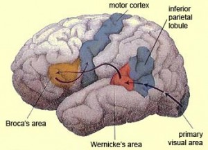 Mirror neurons have also been found in the inferior parietal lobule, where many senses come together—visual, auditory and somatosensory. These specific mirror neurons appear to be only related to the end result of actions such as eating, rather than simpler movement, such as grasping. Therefore, it was thought that these specific mirror neurons were involved in understanding the goals of actions of others, not just the actions. It was considered that these actions of others were matched to previous memories of movements and how they relate to goals.
Mirror Neurons In Humans
 As mentioned above it has been extremely difficult to identify these neurons in humans because of the need for observation of individual neurons in the living. Also, mirror neurons only exist in small numbers near many other types of neurons. Studies of these regions are finding that large numbers of neurons respond in a manner “like a mirror neuron.” This research used evoked responses to see if a large territory and specific actions of mirror neurons are picked up.
Studies in humans have appeared to be similar to monkeys. But, a recent meta analysis of all MRI studies in humans matching action observation and execution found that only a third of the studies touting effects of mirror neuron regions, in fact, included both observation and execution.
One very recent MRI study showed a region of neurons in our brain that appears to ‘mirror’ the space near others, just as if this was the space near ourselves.
Internal Action-Memory Theory
 A current theory is that individuals have a repertoire of performed actions in subconscious action memory that are used in these mirror responses. Therefore, actions similar to those already experienced should have a stronger reaction. This is consistent with the fact that when humans observe animals, the mirror reactions
  occurred with eating, but not with barking. Also, trained dancers and musicians had stronger reactions watching performances than others. Subjects did have stronger reactions with familiar movements than unfamiliar. Training levels correlate with the amount of mirror neuron activation during an observation.
These studies might imply that the observer’s repertoire of experiences of interactions form an internal pool of mental representations.
Is There Internal Emotional Memory?
But, many do not agree with this view, and the research is inconsistent. The implication is that while observing an action, understanding occurs by matching the observation with previous internal movement memories. This view leads to the speculation that this is also true for internal representations of emotions, and the understanding of other’s emotions. Two previous posts described the network of body maps that do exist and are connected with emotions, and the connection of emotions and body maps to our sense of time.
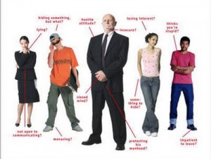 Clearly, observation of social situations involves learning about one’s own emotions and that of others. This internal memory theory would imply that social oriented movements such as facial expressions and body language would also be compared to individual movement memories. MRI evidence seems to imply that the motor actions do relate to imagination of other’s emotions. There has been some imaging showing possible mirror stimulus in the limbic regions.
 Empathy is a very complex subject and is difficult to define. It is critical to emotional responses in social situations. Empathy was demonstrated in mice as well, so many intelligent animals also have it. Empathy, however it is defined, does rely upon shared emotional experiences and also analytical cognitive brain processes of perspective and understanding. In very intimate relationships, the line of demarcation of self and other can become blurred.
The findings related to imitation, facial expression, body language and emotions are not consistent. Studies have used MRI to study imitation and the observation of facial expression with emotions. The premotor cortex appears to show greater stimulation for imitation than observation, but both show somewhat similar activation. This implies that this cortical region might be involved in understanding facial expression of emotion. Similarly, body movement that expresses emotion has also been correlated for both imitation and observation. However, these studies do not have consistent findings for the amygdala and insula, regions critical for emotions (see post on emotions and body states).
 Imitation probably is quite different than spontaneous emotion. In induced emotions, that is pain and disgust, both experiencing and observing include middle cingulate and anterior insula with interior frontal cortex.
Taken as a whole, it is not yet clear how we understand the emotions of others. Although limbic regions appear to have mirror like actions, it does not necessarily mean that mirror neurons are a mechanism for humans to respond to other’s emotions.
Mirror Neurons In Illness
Studies in autism have not been consistent, with some showing less mirror activity and some not. Practitioners of mirror therapy for phantom limb (see previous post on neuroplasticity), where an illusion is created of an active limb in those who have lost a limb, have proposed that this is due to mirror neurons.
 Phantom limb pain often occurs immediately after the surgery, but can occur later, and in most cases pain lasts for many years. Some patients have responded to the mirror therapy. The patient observes a mirror with the illusion that the amputated limb is moving. Then the brain senses a mismatch of the sensation involved. Another variant involves illusory touch that has helped some who didn’t respond to the first. Explaining the neuroplastic changes in this type of therapy through mirror neurons is speculation.
Another type of therapy is called action-observation therapy. A range of studies in stroke patients is using videos of specific movements followed by immediate exercises of the affected limbs. MRI of these patients has shown some improvement. This technique is being tried in Parkinson’s patients, and those with aphasia. These tentatively show that mirror neurons might be involved.
Mirror Neurons Conclusion
 While the current studies cannot confirm the importance of mirror neurons in the basic process of empathy and social understanding, the research is important. The same limitations noted in the “Limits of Current Neuroscience” post apply to all of the research involved with mirror neurons. The only substantial evidence occurs in several animals, not humans. Previous posts have shown that intelligent animals often have quite different brain structures than humans. There might also be very different brain mechanisms involved in understanding others goals, movements and feelings.
While it would be very interesting to have individual neurons that are intelligent enough to have mirror capabilities, conclusions at this time are premature. This would imply a lot of understanding for one cell. Once again, not knowing how mind interacts with individual neurons, brain circuits, and neurochemistry, conclusions about mirror neurons remain speculations.