 The holy grail of many neuroscients is to map neuronal connections and from this explain how the brain (and mind) works. There are approximately 80 billion neurons and there are up to tens of thousands of possible connections from each neuron, which makes the number of synapses an unfathomable astronomical number. In fact, these synapses are part of a very dynamic living cellular process that includes many kinds of glia with elaborate networks across the entire brain and operates over many different scales at the same time. Large projects all over the world are now funded to map the neuronal circuits. The totality of the connections is called the “connectome.” This post will describe the progress made in mapping the connectome thus far, as well as the problems in this approach.
A major problem for these studies is to determine at what scale to study the brain. Mental events affect organelles, synapses, neurons, glia, immune cells, neuronal hubs with local and long distance connectivity, brain regions, whole brain, whole body and interpersonal levels all at once. This post will describe the current research done in the three major scales that are currently being studied. Whole brain studies using fMRI and computer simulations of brain tissue. Medium size studies looking at axonal bundles and connections using brain light microscopes with brain slices. Small studies looking at axons and membranes using electron microscopes with brain slices. Some find that using a centrifuge could help to separate the slices from any liquid to give scientists a better view of the slices.
The details of these three scales and computer simulations will be presented below after a summary of the problems with the entire connectome approach.
The Many Problems with Brain Mapping

A previous post detailed many very serious problems with this approach (See The Limits of Current Neuroscience). This post will give a brief summary of these problems below. First, new problems that have been described since that post.
Since that post was written two years ago, there is more evidence that each individual brain is in fact quite different despite obvious structural similarities. The differences are based on neuroplasticity that produces particular new strengthened circuits based on the unique mental activity of the individual. In fact, a recent study showed that individuals can be detected from their unique fMRI patterns.
Please see the post on the elderly brain (Does Cognitive Ability Improve in Old Age) for a discussion of recent research showing that active elderly brains are, in fact, better than younger brains (despite the prejudice.) This improvement occurs by countless new circuits throughout the brain produced by activity and experience. With these individually unique circuits, they have better pattern recognition and emotional control. The reason this has not been clear before is that elderly do show decreases in word finding. But, in every other way, active brains get better with use, producing many new circuits.The new circuits in the active bright wise elderly include many new connections between the pre frontal cortex (the center of decision-making) and other regions. Other unique circuits are more connections between the right and left sides of the brain.
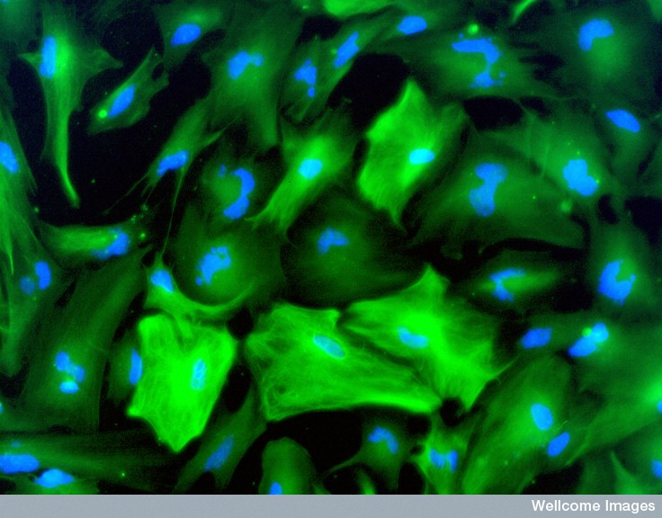 Other recent research emphasized how critical glia cells are in all of the brain’s functions. Astrocytes are very significant in creating, maintaining and pruning all synapses. The astrocyte calcium-signaling network is extremely complex and larger than the neuronal network. The neuronal network depends on the glial network. Also, the resident mobile brain immune cells—microglia—are vital for both protection against microbes, inflammation and infection and, also, responding to mental events that can be as damaging such as stress. Microglia are involved in pruning synapses.
The very complex relationship between the wireless immune system and the wired brain is another huge problem for the brain mapping. Increasingly, the immune and nervous systems have been found to be one system in constant communication for both physical and mental issues (both perceiving and responding to psychological, social and physical stress.) In fact, the immune system is constantly communicating with cytokines to the wired neurons and astrocytes. Microglia work in both systems. Also, the wired neuronal brain is now known to use the same cytokines for wireless communication and is fully capable of producing every type of inflammation through the neuroplasticity process of neuro inflammation. (See posts on neuro-inflammation, choroid plexus, inflammation and depression and stress and unique neuroplasticity).
Summary of Limits of Current Neuroscience
- A widespread experimental assumption in most MRI studies is not certain. This assumption compares an “active” group and control by adding activity to the control’s activity, rather than subtracting.
 Each dot of light on fMRI (voxel) measures average blood flow activity in a region of 80,000 neurons and 4 million synapses over a second.
- Blood flow is determined by astrocytes.
- By the time the blood flow is measured over one second, activity has occurred in milliseconds all over the brain
- Large MRI study used 500 repetitions and found the brain regions chosen in most studies wouldn’t be valid with the small number of repetitions used in most studies. Studies with small “n” find the strongest region. More detailed study showed many small regions all over the brain. The time chosen for the “strongest signal” is arbitrary and many small regions fire before and after time.
- Each test was different because of learning effects from one trial to the next
- The new diffusion MRI techniques measure water, possibly inside and outside the membrane. It is not, yet, clear what this measurement means.
Problems of the “Neural Code”—How neurons decide when to fire and not fire
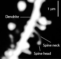
- New synapses are placed in new spatial relationships on the dendrite affecting firing.
- Different types of dendritic spines affect firing
- Many different kinds of neurons (more than a thousand types) fire differently in at least 40 different types of regions with unique properties
- Many different kinds of synapses and post synaptic densities exist throughout the brain
- Postsynaptic densities are different in different regions and each include thousands of various large complex interlocking proteins.
- Individual neurons participate in different neuron subgroup circuits for different tasks.
- Small RNA molecules are transported in exoxome sacs from neurons and picked up, like a virus, by other neurons, even quite far away where they are able to alter function.
- Information transfer between neurons is both hard wired and by wireless cytokine signals and synchronous oscillations.
There Is No center in the Brain for Subjective Experience.
- Detailed study shows most of the brain is used for most mental activities and events.
- There are no modules, but rather many hubs that are very internally connected and very connected to other distant hubs.
- Relation between brain circuits, behavior and subjective experience not really known

Many Other Problems
- Brain wave studies show rolling effects all over brain not one region.
- The “n” of almost all neuroscience studies are too small to find statistical significance
- Changing neuroplasticity produces new synapses and prunes them every day
- Communication is more than axons and synapses—includes other types of communications including brain waves, electrical gradients, vesicles, exosomes and nanotubes
- Brain waves use different frequencies to connect brain regions—‘space’ Information oscillates at 1 to 4 Hz; ‘time’ Information 7 to 10 Hz
- Memory is not a single process but exists in broad circuits around the brain. Very recently, it was found that in a mouse, long-term memory appeared to be in the cortex, not in the hippocampus,
- Extremely complex extracellular matrix is involved in regulating neuronal activity
- Wide range of different neuroplasticity mechanisms occur simultaneously in large brain wide circuits
- Subjective mental experience relates to 12 orders of magnitude of size, from quantum, molecules, cells, organs, brain, society
- Laboratory studies of animal brains show differences in free and captured animals.
- The new myelin code adds to complexity
Current Brain Mapping Studies Using Latest Technology
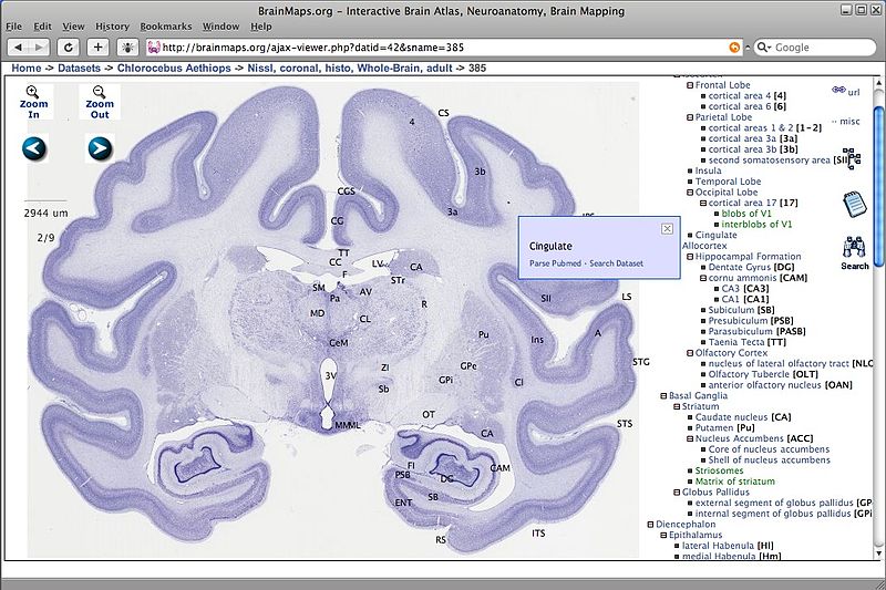
All mapping projects have to consider the many scales of operation that the brain’s architecture encompasses. Synapses and membranes can only be seen with electron microscopes and the whole brain is viewed by fMRI, observing billions of connections at once. The middle scale uses light microscopes and shows axons in circuits.
Connectome studies use the latest and most powerful scientific techniques. MRIs have more and more powerful magnets and new algorithms that use water (diffusion MRI). Other studies use brain tissue slices with more and more powerful microscopes, both light and electron. Strangely, even using these powerful microscopes, human observation is still the gold standard in these studies for disentangling the reams of axons and dendrites in each of the slices.
Large Scale – Whole Brain Study with fMRI

The Developing Human Connectome Project uses a special helmet to scan 1000 babies to show how regions of the brain communicate. The MRI in these studies has the same limitations as others. They hope to understand developing brains.
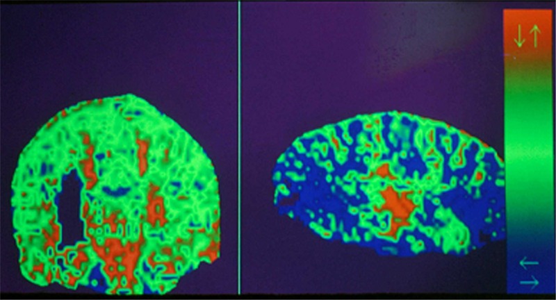
A five-year project funded for 30 million dollars by the United States is called Human Connectome Project. It has been analyzing fMRI scans of 1200 healthy young adults, including twins and other siblings. This study has included tabulating health and lifestyle related information to find any potential correlations. This project is limited by the same fMRI limits described above, although the special MRIs used have some increased resolution with a greater magnet at 3-tesla magnetic. This study looks at blood oxygen levels, while assuming its relation to neuronal activity. The assumption is that areas that are active at the same time are working together (not at all clear since activity occurs in the time scale of milliseconds and the measurements are in seconds.)
Common MRIs use one brain slice picture at a time, whereas this new MRI uses eight images at once. A more recent stronger magnet is 8-tesla and will be more detailed. However as the detail grows, so do the algorithms and assumptions used to build the pictures. These assumptions may or may not be correct.
 Another advance in the MRI helps see the axon bundles clearer. This new “diffusion” technology allows a view of water molecules moving in many more directions than previously (from 64 directions to hundreds). This allows a view of smaller bundles.
Software is used to study the individual differences that occur with the new fMRI techniques and puts them in a view that can be compared between individuals. This same technology is starting to look at children and elderly. With different ages scanned, the hope is to see what occurs at different ages. Additional projects will include specific diseases as well.
Even the directors of these projects explain that these findings would only be approximate.
Middle Scale – Axonal Bundles and Circuits
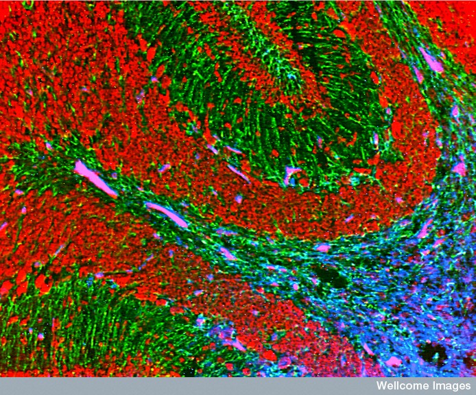
 Brain slices of outgoing axons from a group of neurons can be observed in a very small region. Tagged molecules are injected into mice prior to making the slice of the dead brain tissue to observe communication between regions. This is, then, painstakingly detailed. New research uses marmosets, a small primate.
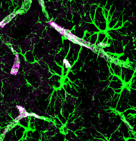
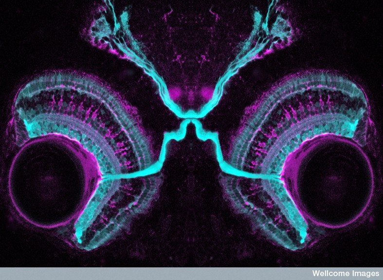
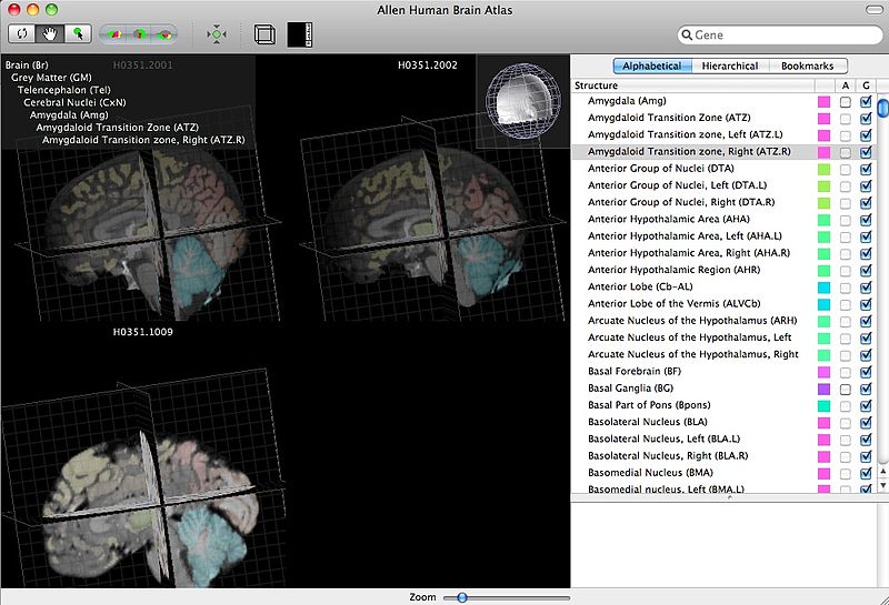
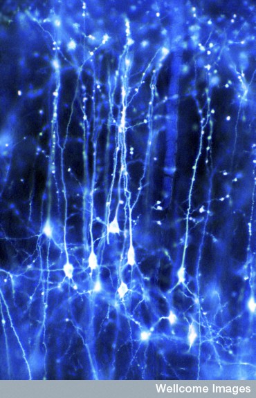
A consortium from the Allen Institute for Brain Science uses living mice. Viruses are injected with a particular gene that produces a glowing protein, which can be observed along pathways between neurons.
A problem with this type of research is the thickness of the slice. If the slice is too thick, then it is very difficult to follow the large number of connections. If it is too thin, then it can affect the axons and produce a poor image. A new special microscope is used with two-photon tomography. By using two photons instead of one the accuracy is increased because the picture is only taken when both photons hit the glowing protein, rather than just one.
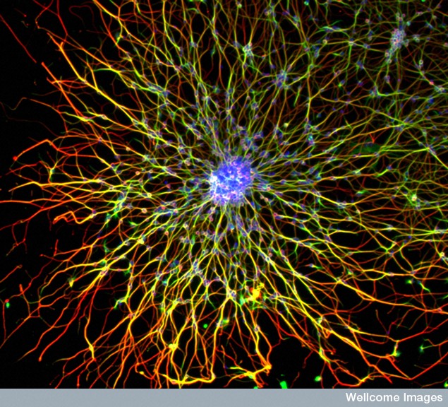
 Another new technique involves placing the brain tissue in a matrix. The picture is taken of the top most visible region before a 100-micrometer slice is made. Each picture is then taken before the slice is made. This technique avoids some of the destruction from the slicing of friable brain tissue. Pictures show 3D tangles of axons that are viewed in 2D pictures. With 100 images, software attempts to untangle the axons. Other projects use multiple colors to help disentangling of the axons.
In fact, software is not very good at visualizing and untangling axons. Much of the analysis is actually done by humans observing the tangles rather than by the software. Having humans visually disentangle the axons is extremely slow.
A project has analyzed 2000 mice brains and is available online to anyone.
Small Scale – Details of the Synapse and Membrane
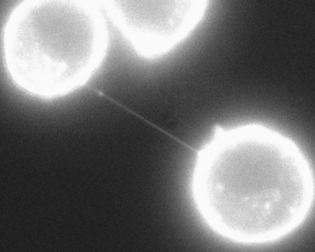
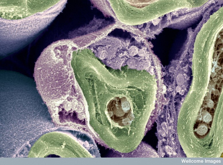
 The smallest scale looks at individual synapses. Electron microscopes give beautiful pictures of cells and synapses. Serial slices are used similar to the mouse slices at medium scale. Small fruit fly brains are scanned before removing an 8-nanometer slice with an ion beam. Each fly brain tissue has 500,000 slices and one brain takes years.
Similar research with mice uses special devices, which are the equivalent of 50 to 100 of these beams at once. Composites are being made where pictures of large slices can zoom in to see detail. As with the middle scale, in fact, software is not good at the untangling and individual visual work by humans is better, but extremely slow.
One group uses a public video game called Brainflight to dis entangle axons. Game players fly along axons through these composite pictures and attempt to untangle the path of individual neurons. (This same type of crowd sourcing has been used with protein folding). The largest product is a mouse retina cube with 1000 neurons and 250,000 synapses. A million of these products would equal a mouse brain.
There is much debate about which level will produce the most useful information. Many think that the middle level is most relevant to describing brain circuits. There is certainly a question of what the fMRI is telling us because of the limitations already described. The smallest views are interesting to describe types of neuroplasticity, but are too small to describe brain circuits that can extend across the entire brain.
Large Supercomputer Brain Simulations
 There are increasing number of projects attempting to make an accurate approximation of neuronal connections in computer simulations to see if this can predict neuronal behavior. Previously a 3D map of a small piece of mouse brain was simulated based on 1000 slices. New much larger expensive simulations are ongoing. The largest European computer simulation project is 100,000 times larger; its first publication was this week.
The large European project funded for a billion dollars has just produced its first major paper with a presentation of a brain simulation. This project constructs a supercomputer simulation of a small region of the mouse brain (0.3 millimeter cubed) in order to study how neuron circuits work. It has been praised by some and, recently, publicly, attacked by 100 neuroscientists in a letter to a journal as a waste of money. It was produced over twenty years with existing data by 82 scientists in 12 institutions.
It simulates 31,000 neurons and their 37 million synapses using 207 different types of brain cells and millions of connections. An algorithm was built predicting how synapses would form and work. The first 1000 neurons were produced by actual recorded data and then the algorithm produced the 31,000 other neurons and their connections. The algorithm predicted locations of 40 million synapses and then calculated pruning.
The project takes models of different kinds of neurons and their shapes; it positions them at different depths. It calculates where they would all touch each other. It first produced 600 million touches. Synapses are given a stimulating or inhibiting character. Five rules of connectivity decide which touches are reasonable and then the algorithm pruned (deletes) the rest. The result after pruning is 37 million touches or synapses.
Experiments with several simulations were performed with these synapses. Possible ion flow through the 37 million synapses was calculated from data for a small number of the neurons. The data was extrapolated to all of the neurons in the simulation. This was then compared with electron microscope data.
 Another simulation studied possible calcium effects. Previous simulations showed bursts similar to sleep (not asynchronous awake behavior). When the simulation calculated calcium for awake animals, it became asynchronous like real brains.
Another simulation was of sensory fibers coming into the cortex such as touch information. They looked for activity and found different patterns, known as “choristers” for firing in groups and “soloists” for firing alone. Neurons were found that fired in three beats or triplets. All of this type of activity has been observed before in the real brain and the simulation identified specific neuron types for the activity.
It is an expensive, interesting project. It is not clear if the assumptions of the algorithm are accurate, nor the results. Each will have to be tested over time. Some scientists feel it is a beginning of something. Others feel nothing is learned and it is an expensive waste. Some problems are obvious even to the directors of the project—no glia, complex neuroplasticity, brain waves, cytokines or blood vessels.
Progress and Problems in Brain Mapping
 The current belief that mind and brain will be deciphered through the neuronal connections is called “neuroncentric” science. While neuronal connections are fascinating and fantastic, they are hardly the entire story. Many posts have been written about the fantastic dynamic levels of activity with astrocytes, microglia, oligodendrocytes and immune cells.
The hope for the connectome researchers is that mind is a creation of brain, which is thought to be a computer of some type. The extreme view is that mind doesn’t exist at all, but is an illusion—an epiphenomenon—much like a digital character in an animation created by the computer. It is spurred by the philosophy that there is no mind and no meaning in the universe, but rather only random molecules bumping into each other and creating an illusion of mind through physical forces.
Current research doesn’t support this view; the deluge of new science described in these posts supports the notion that mind exists in cells, organelles, and molecules. The latest and best current science shows a large amount of evidence consistent with the new paradigm that mind exists as an integral part of nature like matter and forms of energy. While the work being done to map the brain is extremely valuable and interesting, it is necessary to point out dramatic and potentially insurmountable obstacles in this quest to reduce mind to neural connections.