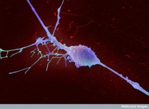
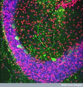
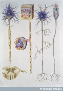
When neurons differentiate from stem cells, they become a very particular type of cell that can last a hundred years. The post How Many Different Types of Neurons are There noted that there are at least a thousand very different species of neurons with varied structures and functions. How does the cell know how to become this exact type of cell? Equally important is how can it maintain this identity for such a long period of time. It has been found recently, that this identity must be carefully sustained through active work of the cell using very specific genetic networks. Without this very specific constant daily genetic stimulation, it will not stay as that type of neuron for even a day.
Maintaining identity is another characteristic of cellular intelligence, which has been described in many previous posts. Individual cells show a surprising array of intelligent behaviors that are hard to explain. Cells are able to signal back and forth to each other for many purposes even at great distance and to totally different kinds of cells. Cells know exactly where they are in the body and in the brain. They can travel long distances along varied terrain with many different techniques to arrive at a very specific place. They know how large they should be. Now, we describe the fact that they choose the type of neuronal identity they should have and actively reinforce this each day. Maintaining neural identity is a complex but lifelong activity.
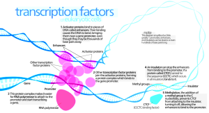
One of the many regulatory mechanisms of genetic networks utilizes large proteins that bind to very particular places on the DNA and stimulate transcription—the process of making messenger RNA that will be edited and then manufacture proteins. These protein molecules that stimulate the genetic process at particular places along the genome are called transcription factors (TFs). TFs are one of the major ways that neurons form their particular identity and stay that way. But, the process is very complex and different for each type of neuron and neuronal behavior. In addition, there are complex feedback loops involved and other epigenetic factors that shape the chromatin to maintain these particular TFs. These genetic mechanism are critical for maintaining neuronal identity.
Cellular Intelligence
 Previous posts have described aspects of the remarkable intelligence of cells. Neurons, which are extremely specialized and complex, show even greater intelligence than many cells.
Cells use a very complex language for communication. Cellular signaling uses many different modalities—cytokines, chemokines, neurotransmitters, electrical signals, photon signals, nanotube signals, and vesicles filled with genetic information. The individual cell is able to integrate the messages from all of these sources and send back specific signals.
Cells are able to know where they are in relation to organs and other cells without GPS. This ability is surprising since it lives in a world of billions of cells. It is equivalent to a human knowing what country they live in and how this relates to other countries. In fact, they perform complex calculations using the relation to other cells, gradients and specific signals.
They can also, travel to very specific places that are quite far away, such as leukocytes finding an infection or trauma in another part of the body, neurons finding precise locations in the developing brain, and axons traveling to one end of the brain to the other. Cells in limbs and organs know precisely how they form the proper shape and when to stop its expansion. To travel, cells change their shape and their locomotion mechanisms. They signal for advice.
To know their place, cells use multiple measurements and rules to calculate their location and they adjust for many sources of error in these calculations. They check measurements with neighbors and use feedback loops, timed measurement and gene regulation. Each situation can involve many different variables that can be altered with temperature, metabolism and other environmental factors. It is quite remarkable that individual cells are mathematicians.
Another intelligent characteristic of cells is that they know what exact size they should be. This process is, actually, quite complex. Size is determined by many factors—synthesis and breakdown, nutrition, environmental factors, protein production and autophagy. In fact, it is quite complex involving the specific functions and physiology of the cell and the organ. They way the stem cell differentiates to a specific sized cell is important as well.
It turns out that knowing what type of cell a neuron should be and maintaining this identity is really quite complicated.
Many Neuron Types
Determining the species of animals is difficult because genetic similarities and differences may be quite distinct from appearance characteristics. Attempting to decipher different neuron species encounters the same difficulty.
There has been no clear way to define how many distinct terminally differentiated neurons there are. But, now species are being defined with a combination of specific function and structure along with genetic makeup. Appearance includes structural differences in shape and positioning of dendrites and axons. Many other functional characteristics define these differences—architecture, density, size, electrical properties, microtubule scaffolding structures, synaptic structures and circuits. Dendrites have different spatial and timing arrangements. It is clear at the present time that there are at least 1000 different distinct types in at least 40 different regions.
Neuron Identity
The specific type of neuron is first expressed in the fetus by a very particular network of genes, sometimes called “gene batteries”. The function of these gene batteries then must be maintained for up to a hundred years with very exact regulation, sometimes using the same transcription factors that first start functioning in the fetus, sometimes new ones. If this identity is disrupted then psychiatric and degenerative brain diseases can occur.
It is quite remarkable that neurons can form so many distinct types that maintain their shapes and functions for decades, while at the same time being extremely adaptive and changeable with a vast array of neuroplasticity mechanisms. Neurons are constantly changing their shape by growing and pruning axons, dendrites and synapses. These changes include very complex alterations of scaffolding molecules (see post on The Enormous Complexity of Transport Along the Axon). Most neurotransmitters that are used are usually stable throughout the years of a neuron’s life, while some neurons, in fact, alter the neurotransmitter (discussed later and in a previous post, Neuroplasticity Involving Changes of Neurotransmitters) . Each neuron, also, takes part in very distinctive circuits. Those factors that maintain the identity of the neuron must maintain the use of neurotransmitters, the shape and the particular circuits.
Study of a worm shows 120 types of different neuron types defined by their form, their circuits and their functions. Their line of descent from other cells, also, defines them. Special genes respond to specific types of molecules utilized by the particular neuron type. These include receptors, enzymes that make neurotransmitters, the molecules that transport the neurotransmitters, specific ion channels (see post on Fantastic Complexity in Brain Potassium Channels) and other factors specific to the function of the neuron.
Certain transcription factors (TFs) trigger the division of cells to make the new cells and what type of neuron they become. In the worm, more than half of the 120 types have specific different TFs using cis elements. Groups of TFs are coordinated in different ways to differentiate the cell; these are called “terminal selectors.” In experiments where these TFs are deleted, many of the specific aspects of the neuron disappear, such as receptors and ion channels. But, the more common shared functions such as making vesicles do not. Studies of the worm show that these same TFs need to be maintained for the life of worm to continue these characteristics. Examples include specific glutamate sensory neurons, cholinergic motor neurons, and all dopamine neurons.
Studies show epigenetic histone modifications of chromatin structures are not sufficient to continue this differentiation, but, also, need the continued daily production of the protein TFs.
Vertebrate Neurons
In vertebrates, the groups of TFs are vital for all of the functions of neurotransmission including manufacture of all major neurotransmitters and their particular transport proteins. Large arrays of TFs maintain all of the different neuronal characteristics throughout the brain at the same time to maintain all of the different neurotransmitters in circuits. They maintain the synthesis, the creation of the special vesicles that are released and the particular uptake molecules. These TF patterns start early and continue throughout adulthood. If the TFs are removed, then the neurotransmitter functions stop. The gene batteries are gone within days. This occurs for serotonin, dopamine, acetylcholine and noradrenergic neurons.
What is quite remarkable is all of these different neurotransmitters have different groups of TFs for each possible function. The research has been quite complex to uncover all of these details. Some of these TF patterns have other elaborate functions, such as maintaining the functions of mitochondria and oxidation. For many situations, the TFs affect the rate-limiting step in the manufacture of critical proteins.
There are some neurons who are able to do without the particular TFs from fetal development. They use new patterns in adulthood. This was found is particular for serotonin neurons. In these cases, the types of TFs evolve, but still keep the same identity of the neuron. In other situations, TF groups and patterns take over the function of another type of neuron and some work together to maintain a particular function they have taken over.
Very Complex Maintenance
 Neurons developed during the fetus mature and produce many different signaling and molecular pathways. The exact pathways from the fetus are usually greatly decreased in the adult and mature patterns take over. Still, they maintain genetic regulation of TFs from this early period despite a very different situation and metabolic stream.
To maintain these fetal TFs, a form of auto regulation occurs, where feedback loops of different types keep the TFs going. In one example, touch sensory neurons require a particular set of genetic networks and particular proteins that create the differentiation into the particular type of neuron. Some of the original patterns join together with another to modulate, regulate and maintain the function of the neuron once the differentiation is over. Special loops control the genes and these are extremely particular to the exact type of neuron. It is not clear how these complex feedback loops develop from original fetal genetic programs. But, the loops can be extremely complicated and they have only been worked out for several types of neurons.
Adult Neuron Complexity
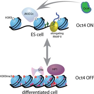
The original stem cell creates the new neuron and then particular genetic patterns are temporarily triggered. Once the type of neuron is created, feedback loops are triggered to maintain the situation using many other pathways. These patterns activate the particular development of all of the TFs needed to make the precise neurotransmitters, receptors, transport molecules, vesicles, ion channels and the thousands of different molecules that hold the synapse together including the specific and highly complex post synaptic density. The TFs are regulated to maintain the complexity of the particular neuron and the exact types of axons, dendrites and synapses.
There is great room for error in such a complex process. Experiments that alter some of these feedback loops create behavioral changes in animal experiments that appear to be similar to those seen in psychiatric and brain neurodegenerative diseases.
The feedback loops are highly complex and very specific, even including particular cytokines. As well, factors alter the shape of the chromatin (see post on Vast Complexity of Chromatin 3D Shapes), but are dependent on the particular TFs. There are examples of where the TFs that created the original connections in the fetus and young child, are necessary to maintain it.
One example of the need to keep TFs occurs in vision to avoid degeneration of the light receptors. One feedback loop continues the mechanism whereby the proper amount of rods and cones are created and maintained. Without these constant regulated particular TFs there are too many rods or cones. But, there are thousands of other examples.
Creation of Neuron Identities
 Recent research shows that while some of the original fetal TF patterns were critical to maintain neuron individuality and function, there are, also, special new patterns that appear to maintain the identities over time. In fact, genetic networks that create the differentiation, trigger totally new programs that are necessary to maintain the identity. There are many examples now of these dedicated systems of TFs that are unique, start in the adult brain and are absolutely necessary for the identity of the neuron.
An example is dopamine neurons that have a particular program to create the neuron type and then totally different ones triggered to maintain them. This latter genetic network is tied to the mechanism of apoptosis and avoids programmed cell death that would occur without them. The dopamine TFs are part of the protection of the neuronal circuit as well and have been implicated in the causes of Parkinson’s disease.
Serotonin neurons, also, have been found to have a particular set of totally new maintenance TFs. Without these, the raphe nucleus has a gradual loss of identity as the main brain serotonin source. They are necessary to avoid apoptosis. These factors appear to be necessary to attract other factors that maintain the neuron.
What is unusual about these TFs is that there are a wide range of different genes and factors for each type of neuron that are necessary to maintain identity.
Plasticity

Another unusual feature of these genetic networks is that they are flexible and can change. TFs need specific kinds of activity to start them and then feedback loops of this activity to maintain them. These plasticity changes occur in response to the environment of the neuron. The genetic programs are created first by the fetal gradients that produce specific activity in regions of the fetus. The mature new TFs are somehow triggered as the neuron matures. And then, these are altered by specific triggers later in their function. These alterations can change the identities of some neurons and trigger the production of different types. Is it possible that the activity of the synapses themselves produce feedback loops that alter the identity of neurons?
Sensory input or other environmental factors can cause the TFs to change. One example of this switch was described in a previous post on neuroplasticity. In the hypothalamus, specific inhibitory interneurons stop functioning as dopamine neurons and instead change to become somatostatin neurons. Recent research shows that this is triggered by specific mRNAs, which make particular TFs. Therefore the mechanism of the TFs altered the neuron identity for a particular response.
Another switch occurs from adrenergic to cholinergic for particular hormone glands. This is triggered by TFs that produce cytokines that trigger the adrenergic neurons to change. Both of these different neuron identities once established are maintained through unique and specific genetic patterns. There is some evidence that part of this mechanism involves changing the structure of the chromatin.
Alterations in Chromatin
Very particular sequences of proteins bind to DNA and stimulate the cell to become one type of neuron. These sequences and others that are triggered are necessary to constantly maintain that structure and function of the neuron. Without the constant triggering of these programs the neuron will deteriorate immediately. Epigenetic markings on histones are, also, necessary to maintain these mature states. These tags mark the active regions that TFs are stimulating. However, these markings alone will not maintain the neuron. Both are necessary.
There are now many examples of how if these patterns of transcription proteins are altered then neuropsychiatric disorders can occur. This can occur with anxiety disorders and Parkinson’s.
Maintaining Neuronal Identity
Cells know where they are, where they need to go among billions of other cells. They communicate back and forth with many different cells simultaneously. They know what size they should be and what type of neuron among hundreds of billions of other neurons They maintain this identity for a hundred years. When the particular neuron is created by the stem cell particular TFs are used. Later, others appear to continue the same identity. But, then through neuroplasticity these can change. Where is the direction for all of this?
How can an individual cell be so intelligent? How can an individual cell integrate so many different kinds of information and then act in concert with large numbers of other cells? It must maintain a daily set of specific genetic triggers for up to a hundred years?
Where does this intelligence lie? Where is the direction? Isn’t it most reasonable to presume that all of these cells are tied to a source of integrated information or mind?
