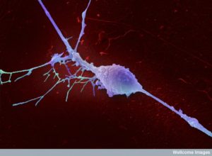
 As technology advances, we are able to observe the behavior, decision-making, and communication of individual cells. This complicates understanding how activity from individual cells is integrated into the function of organs and organisms at very different scales. A recent set of posts described the new findings on cellular clocks that show each individual cell has its own clock. In fact, each different type of cell in each organ has its own unique clock along with a fundamental genetic regulated structure that is the core of all cellular clocks. These posts described the current research attempting to find out these individual cellular clocks integrated with metabolism and immune responses in tissues and brain clocks that synchronize many functions across the entire human physiology.
There is no more complex environment than the brain for these questions as to how individual cell decisions integrates into global cognitive processes. Recent research shows definite behavior of individual neurons, such as the “Jennifer Anniston neuron” that was seen when deep electrodes stimulated specific individual cells. No one knows how this information is translated into human brain activity, but it appears to be related. More recent evidence questions possible structural constraints on different cells might determine how decisions from individual cells might be utilized.
This post addresses what is known about the lifestyle of single neurons and how this might be relevant to human size events such as cognition, sleep, seizures, and sensory data.
Brain Theories and Measurement of Brain Activity
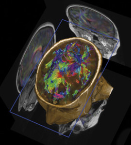 Most theories as to how the brain works involve collective activity of large numbers of neurons in circuits or production of electrical oscillations that together form synchronous brain waves. Possibly, this is because research could, until recently, only describe these larger structures and activities. Current advances for the first time allow the beginning of the study of individual neurons. Since many different kinds of cells demonstrate intelligent signaling and activity, it is crucial that we understand perhaps the most complex and intelligent cell—the individual neuron.
MRI imaging devices have been considered the gold standard of observing brain activity. What is not widely understood is that there are severe limitations on the interpretation of MRI’s. In fact, top national brain experts acknowledge that all of the statements correlating human mental states with particular regions are speculative. To understand how this could be true, please read the previous posts on the limitations of MRIs and the limitations of current neuroscience.
 MRIs measure the activity of a small regions of the brain averaged over seconds and each measurement produces a dot of light on the computer screen. This dot of light is called a voxel (like a pixel on a regular computer) and represents the blood flow in a region of about a 100,000 neurons.
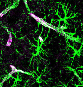
In fact, they don’t measure neurons at all, but rather the blood flow in that region, which is controlled by astrocytes with end feet completely surrounding blood vessels. Astrocytes open and close the small blood vessels controlling how much oxygen neurons need. This blood flow measurement doesn’t produce an exact correlation to neuronal activity, but approximate. Recent studies showed that MRI measurements might be after or before the actual activity of neurons making precise locations impossible.
The other issue is that neuronal circuit activity occurs in milliseconds, whereas the MRI averages signals in that small region over seconds, a thousand times slower. Another problem in interpretation is that individual neurons can be part of one circuit one moment and another the next millisecond and this cannot be picked up.
Individual Neurons
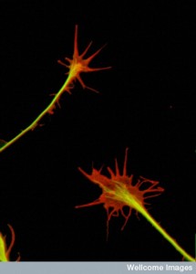 The main description of individual neurons up until recently has been for peripheral nerves. Most diseases are described in terms of the activity of a large group of neurons. Neurodegenerative diseases and demyelinating diseases have pathology related to many neurons. Stroke and trauma and infections relate, also, to many neurons at a time.
Today, many new technologies are merging to allow some observation of individual neurons. Research shows that in non human primates simple movements can be correlated with activity from small groups of neurons. One of these experiments controlled a computer cursor. These patterns of activity are able to be understood and then messages from this small number of neurons able to maneuver prosthetic limbs. Research showed that individual neurons understood some of the movements.
Movement and the Individual Neuron
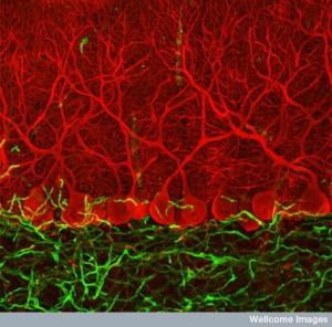 Individual neuron measurements have been used to place electrodes in the basal ganglia movement centers to attempt treatment of Parkinson’s with deep brain stimulation. The electrode is placed as accurately as it can be in the sub thalamic nuclei (STN) or the globus pallidus. The activity measured by the tip of the electrode changes as it is moved into place. Patients are awake during this activity and can comment on their experiences as the electrode measures different neurons.
These electrodes are also used as treatment for difficult to treat tremors and dystonia. Individual neurons in the basal ganglia and motor thalamus have been measured with specific electrical activity patterns related to the particular tremor. The individual neuron activity is then either part of or stimulating larger groups of neurons firing at the same rate. Ordinarily, many smaller circuits are separated, but in these tremor diseases the separation breaks down to allow transmission to a larger area of a particular type of tremor activity.
Seizures and the Individual Neuron
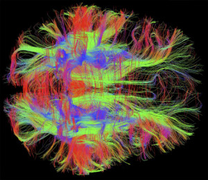 While the purpose of one type research is to connect human prosthetics, it, also, advances measurement of individual neurons. Movement has been easier to translate from animal brains to humans since the M1 movement region is similar in non-human primates.
But, with seizures, there is no specific correspondence. Somewhat by chance, invasive research on animals identified a seizure focus. Specific neurons with increased activity were identified in the cortex and in the amygdala and hippocampus that correlated with seizures. Most of the findings into auras and subclinical activity related to seizures.
Measurements of single neurons with microelectrodes studied high frequency brain waves. It showed differences in the electrical activity of individual neurons on the two sides of the brain, with greater activity on the side where the seizure occurred.
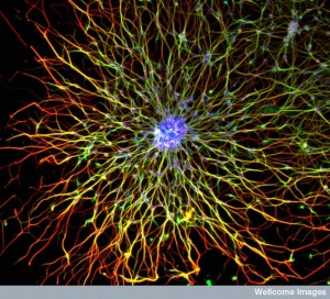 Another study measured local field potential and individual neuron activity. The activity was measured in a single cortical column of neurons. This study identified differences in various layers—mainly greater activity in layers IV and V.
A different technique measures many different individual neurons. They found a very complex pattern of different responses during seizures and surrounding the seizures in different types of neurons.
It showed that there are complex interactions between different kinds of neurons, mainly in regions not in the primary focus but nearby regions where the activity was spreading. There appears to be neurons that are excessively synchronous, but others that have different behavior. This research implies that seizures are not just an excessive activity in all neurons, but rather a complex interplay between different kinds of neurons and glial cells. Also, there were findings in individual neurons minutes before the onset of the seizures.
Anesthesia and Individual Neurons
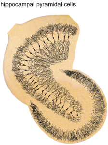 It is surprising how little is known about the critical process of anesthesia used for surgery. The medications used have particular effects on receptors, but the basic way that the brain is quieted is not at all clear. There are particular EEG patterns that arise, but it is not clear exactly how these low frequency, high power, gamma and alpha rhythms arise.
Recently, measurements of individual neurons showed that they can produce slow oscillations. The individual neuron becomes disconnected from distant cortical oscillations and instead is locked at a particular low frequency. Whatever is occurring in the individual neuron has separated it from the rest of the cortex causing disruption of consciousness.
Cognition and the Individual Neuron
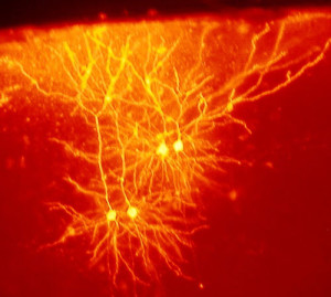 During deep brain electrode placement, studies have been done on memory, behavior, vision, hearing, reward and language. Some of these studies show that individual neurons in the higher regions of the cortex appear to be related to complex topics, such as the famous “Jennifer Aniston Neuron”.
There do appear to be specific functions that individual neurons take on, among many possibilities. An example is discussed in a previous post where specific CA1 hippocampal neurons can be either time cells (measuring intervals of events in a memory) or place cells (measuring locations) or both. They can, also, be related to particular activities at those times and places. Other studies find individual neurons correlating with specific activities and motivations. Human faces have been noted to be the individual neuron focus in some of these studies.
In the most recent study, individual neurons in the amygdala responded mostly to seeing an entire face. The cells don’t respond well to a face with one small part removed. In fact, the more of the face that is removed, the more they respond, opposite to what we would suppose. These neurons respond to the totality of a face. The brain doesn’t appear to like anything to be wrong with that face, such as missing pieces.
Sleep and the Individual Neuron
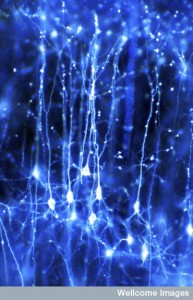 Another finding comes from recent research into sleep and shows that some neurons fall asleep while others are awake. As the percentage of neurons that are asleep increases, we experience “sleepiness” as an organism.
There are general global brain waves that occur in sleep. But, recent findings show that there is great variability in the state of individual neurons, even while many neurons have created a generalized larger state of oscillation frequencies. Studies have clearly shown a great effect on memory and learning during phases of sleep. Memory consolidation is definitely increased during sleep. This means that multiple different processes are going on at the same time, involving either smaller numbers of neurons or individual neurons.
Sensory Neuron and the Individual Neuron
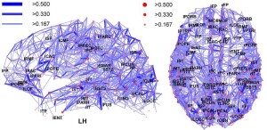 How do we respond so specifically to a sensory input such as a comment from someone else? Do whole regions respond or do individual neurons have a role? It does appear that individual neurons can deal with advanced concepts and at least parts of perceptions. But, is that how it works?
Measurements in monkeys who are given incomplete information, show specific cortex neurons correlated with responses. In a recent study, neurons in the visual cortex fired for particular sensory stimuli, but responded stronger when closer to the ideal stimuli and less if it is almost the proper stimuli. This represents a vote from individual neurons for a particular experience occurring. The study showed that neuron’s individual rates will vary with other neurons that are measuring other parts of the visual stimuli (lateral versus vertical movement). It is now, also, known that expectation plays a role with top down input modulating and evaluating the visual information. The individual neuron has a complex relationship to the eventual understanding of the perception and then actions taken.
Single Neurons Detect a Sequence of Events
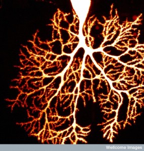 Even more recent research shows for the first time that single neurons can detect and respond to a sequence of events. Surprisingly, they perform this functions very well. But, even more was discovered. Individual dendrites also can distinguish these sequences. Dendrites are described in previous posts and are noted to be very tiny. Each neuron can have tens of thousands of dendrites and each makes decisions that are then somehow merged into decisions for the entire neuron about whether to fire or not and what type of firing will occur.
This sequence experiment was done on a mouse, where dendrites were visualized with fluorescent dye. A laser activated a tiny spot on the individual dendrite. Each different sequence on the dendrite had distinct responses. The individual dendrite was able to accurately understand sequences.
The Final Decision of An Animal

But, the larger question remains as to how these individual dendrites and neuron outputs are used by the circuit and the brain as a whole. These findings are considerably different than sequences needing a group of neurons working in order and in a circuit. Even more unusual is the fact that (even young childrens’) brains are able to analyze and respond to information that is, in fact, so complex that the most advanced super computers cannot. Can individual cells do this as well?
Another new set of research shows that in a monkey brain, these responses of individual neurons are correlated somewhat with the final decision of the animal. This research used very limited visual information and showed that the final decisions of the animal using billions of neurons was perhaps relevant even to this small amount of information input given to individual cells.
It could be that the local neuron responded to the decision that was made by the larger circuits and brain. But, it doesn’t answer the question as to how the individual neuron relates to the brain.
One theory proposed is that each decision of individual neurons is restricted in its output. This could possibly mean that all kinds of information are coded, and responded to, but are only used in certain circumstances. How this happens is not known.
What Do Single Neurons Know
 Current research supports the notion that all cells communicate with a similar language and that decisions are made in conversations between a wide range of cells. One example of this are the discussions in the gut among lining cells, immune cells, and microbes that determine digestion and the responses to the varied microbes.
Recent research also shows that neurons have wide ranging communication among cells not just with neurons, but also with glial cells, immune and blood cells, and tissue cells. Even though decisions are made in this wider context, individual cells appear to play a role.
How does the intelligent decision making and conversations of individual cells translate into the function of organs, tissues, and creatures?